Endosonography with 25-gauge core needles for immunohistochemistry and molecular testing in lung cancer patients: a feasibility study


 CC BY-NC-SA 4.0
Endosonography with 25-gauge core needles for immunohistochemistry and molecular testing in lung cancer patients: a feasibility study
Introduction
Lung cancer (LC) remains the most common cause of cancer‑related mortality worldwide. Despite the efforts made to detect it early, only 15% to 20% of newly diagnosed LC patients are in early stages at the time of the diagnosis and are eligible for surgical treatment. A targeted oncological treatment should be considered in the majority of patients with advanced LC (stage IIIB and IV). It is important to obtain from the lesions adequate cytological or histological samples that are suitable for immunohistochemistry (IHC) and broader molecular testing (MT).1
A combination of positron emission tomography with computed tomography and minimally invasive procedures including endobronchial ultrasound‑guided transbronchial needle aspiration (EBUS‑TBNA) and endoscopic ultrasound‑guided fine‑needle aspiration (EUS‑b‑FNA) with single EBUS scope is a standard procedure in LC staging.2 Recent guidelines on the technical aspects of EBUS‑TBNA recommend using a 22‑gauge (G) or 21G cytological needle.3
For decades, transthoracic and transabdominal core needle biopsies have been the standard procedures for the diagnostic workup of malignancies.4 Both thicker cytological 19G needles and core histological 22G/25G needles proved to be useful in performing core EUS‑guided biopsies in gastroenterology5 (see also references 1 and 2 in Supplementary material). Thereafter, histological core needles started to be used endobronchially, but their efficacy to obtain sufficient material for IHC and MT in the diagnostic workup of patients with LC is still unknown.
In some cases, it is crucial to assess the histological structure of the lesion to diagnose and treat the patient correctly (eg, lymphoma). Core needle biopsies can sometimes provide such a sample, but if EBUS‑TBNA is not conclusive, mediastinoscopy is still a confirmatory test.6
The aim of this study was to evaluate efficacy of EBUS/EUS‑b‑NA using histological 25G core needles in a detailed diagnosis of patients with advanced LC.
Patients and methods
A single‑center prospective observational study was conducted in John Paul II Hospital in Kraków, Poland. Consecutive patients with confirmed or suspected advanced LC were included. Since the procedure is routinely used, the approval of bioethics committee was not required.
After the informed consent was obtained, the EBUS/EUS‑b‑NA procedure was performed under mild conscious sedation with intravenous midazolam (2–5 mg) and fentanyl (0.025–0.1 mg). Flexible videobronchoscopes with a integrated convex ultrasound probe (BF‑UC180F, Olympus Medical Systems Corporation, Tokyo, Japan), which can visualize mediastinal and hilar structures, were used for all procedures. The biopsies were performed with 25G core needles (EchoTip ProCore Endobronchial HD Biopsy Needle, Cook Endoscopy Inc., Limeric, Ireland) with 2 to 5 needle passes in at least 2 selected areas. Both the smears and the cell blocks were prepared from obtained samples.
If the biopsy specimen was positive for LC, it was sent to perform IHC tests: thyroid transcription factor 1 (TTF‑1), p40, CD56, Ki67. If non–small cell lung cancer was diagnosed, the specimen was sent to run molecular testing for mutations of epidermal growth factor receptor, translocations of anaplastic lymphoma kinase, and expression of programmed death ligand 1.
If the biopsy result was negative or inconclusive, it was repeated with standard 22G cytological needles as the confirmatory test.
Results
From January 2019 to April 2019, 20 patients (13 men [65%] and 7 women [35%]) at a mean (SD) age 68 (6.4) years underwent endosonography. Histological examination was conclusive in 18 patients: small cell lung cancer (8 patients), adenocarcinoma (6 patients), squamous cell carcinoma (3 patients), and not otherwise specified (1 patient). Sensitivity, accuracy, and positive predictive value (PPV) for the histology and IHC tests in the whole group were 90%, 90%, 100%, respectively. In 2 samples with false‑negative results, in which some undifferentiated cancer cells were only found, EBUS‑TBNA with a 22G needle confirmed small cell lung cancer (1 patient) and squamous cell carcinoma (1 patient).
The mean (range) size of biopsied lymph nodes was 30.25 mm (17–70 mm). EBUS‑TBNA was performed in 10 patients with LC, and EUS‑b‑FNA in 7, as a single procedure. Combined procedure EBUS/EUS‑b‑NA was used in the next 3 cases. The nodal station 7 was biopsied in 13 patients. Stations 4R, 11L, 4L, 10R, 2R, and 8 were biopsied in 8, 5, 4, 3, 2, and 2 patients, respectively. On average, 2 different nodal stations per patient were biopsied.
In 8 of 11 patients with non–small cell lung cancer sensitivity, accuracy, and PPV for endosonography with 25G core needles for MT were 72.7%, 72.7%, 100%, respectively. No complications after all biopsies were observed.
Discussion
Endosonography with 25G core needles in our prospective observational study had high diagnostic yield for histology, IHC, and MT and good safety profile in patients with advanced LC.
At present, there is a variety of cytological needles available for biopsy: thicker 19G, standard 21G and 22G, and thinner 25G.7
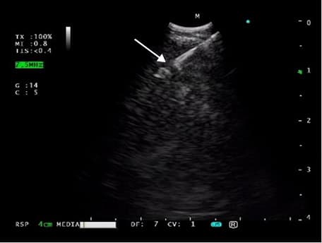
The EchoTip 25G ProCore needle was designed to provide a histological biopsy material, because it is equipped with a core trap close to its tip (Figure 1). It is also 33% more flexible compared with the standard 22G needle. A maximum extension of this needle is 5 cm.8 Nevertheless, the literature describing usefulness of this needle in obtaining material suitable for IHC and MT is scarce.
Our study showed that 25G ProCore needles can be successfully used for this purpose. Those results must be considered as preliminary and requiring further investigation, because our study had its limitations, namely, a small group of patients and a noncomparative protocol.
Also considering statistical analysis, both accuracy and PPV are influenced by a selection bias, so their value is limited.
There is only one retrospective review comparing core biopsy needles (EchoTip 22/25G ProCore) with standard ones (SonoTip II 22G) in which a yield obtained by the first one was improved comparing with the standard one in testing for malignancy.9 On the other hand, more recent data do not show significant difference between 22G and 25G cytological needles regarding specimen adequacy for histology and molecular testing.10,11
Also, the comparison between 22G and 25G ProCore needles for mediastinal and hilar lymphadenopathy presented by Yang et al12 did not show significant difference in the diagnostic yield.
As the core needles are significantly more expensive, and their diagnostic yield seems to be of no advantage comparing with standard ones, it is necessary to identify the precise indications for the use of 25G ProCore in clinical practice. The data from the literature is very limited (see references 3 and 4 in Supplementary material). However, both conclusions drawn by other authors and our experience point to improvement of flexibility of EBUS scope loaded with a 25G needle, which is important in puncturing both the upper paratracheal region and peripheral hilar lesions/lymph nodes (eg, level 12L in the left upper lobe). The other important indication for a 25G needle could be the need to obtain a biopsy specimen from a lesion located behind a vessel (transvascular biopsy) or hypervascular tumor (ie, metastatic renal cell carcinoma).
Both flexibility and sharpness of 25G needles that allow for more precise puncturing of small or fibrotic (after induction therapy) lymph nodes make them useful also in LC (re)staging. But it is under debate whether the more expensive 25G ProCore histological needles have any advantage over 25G cytological ones in this indication.
In conclusion, the endosonography with histological 25G core needles is feasible and safe, so it seems to be a reasonable approach in selected clinical situations for IHC and MT in patients with advanced LC.
- Planchard D, Popat S, Kerr K, et al. Metastatic non‑small cell lung cancer: ESMO Clinical Practice Guidelines for diagnosis, treatment and follow‑up. Ann Oncol. 2018; 29 (suppl 4): iv192‑iv237. | Crossref
- Vilmann P, Clementsen PF, Colella S, et al. Combined endobronchial and esophageal endosonography for the diagnosis and staging of lung cancer: European Society of Gastrointestinal Endoscopy (ESGE) Guideline, in cooperation with the European Respiratory Society (ERS) and the European Society of Thoracic Surgeons (ESTS). Endoscopy. 2015; 47: 545‑559. | Crossref
- Wahidi MM, Herth F, Yasufuku K, et al. Technical aspects of endobronchial ultrasound‑guided transbronchial needle aspiration: CHEST guideline and expert panel report. Chest. 2016; 149: 816‑835. | Crossref
- Babińska A, Berendt‑Obołończyk M, Śledziński Z, Sworczak K. Liver metastasis as the first manifestation of differentiated thyroid cancer. Pol Arch Intern Med. 2018; 128: 478‑479.
- Witt BL, Adler DG, Hilden K, et al. A comparative needle study: EUS‑FNA procedures using the HD ProCore(™) and EchoTip(®) 22‑gauge needle types. Diagn Cytopathol. 2013; 41: 1069‑1074. | Crossref
SUPPLEMENTARY MATERIAL
ARTICLE INFORMATION