Introduction

The hemostatic system represents a delicate equilibrium between procoagulant and anticoagulant elements. Physiologically, 3 main anticoagulant pathways dominate in this equilibrium and enable blood flow through the vascular system: tissue factor inhibitor, protein C, and antithrombin pathways. Any distortion in the function of these anticoagulants will definitively increase the risk of a thrombotic event through an inefficient control of procoagulant reactions. This review focuses on probably the main endogenous anticoagulant, antithrombin, and the clinical consequences of its deficiency. The aim of the present article is to describe this protein and the disorder as well as to provide useful guidance for its proper diagnosis and management.

Antithrombin molecule

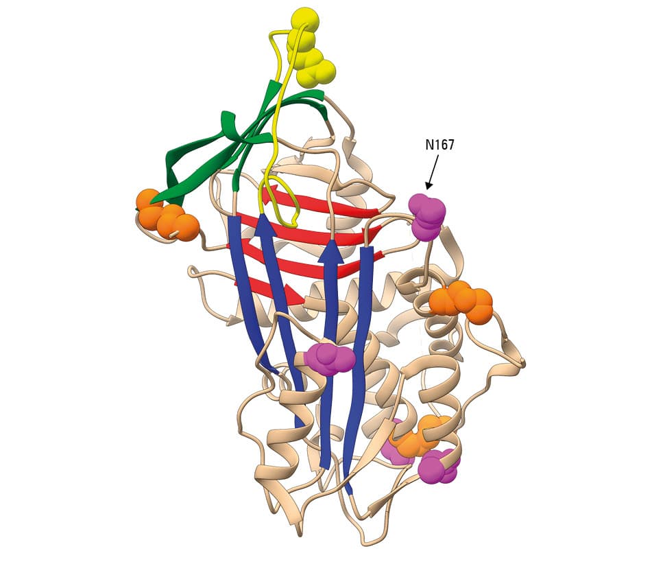
Antithrombin is a glycoprotein, a product of hepatic synthesis. The signal peptide of 32 amino acids guides the nascent protein to the endoplasmic reticulum, where the mature protein of 432 amino acids is subjected to 2 post-translational modifications: N-glycosylation at 4 asparagines (Asn128, Asn167, Asn187, and Asn224) and 3 cysteine disulfide bonds (Cys40–Cys160, Cys53–Cys127, and Cys279–Cys462) (figure 1). Since Asn167 is inefficiently glycosylated, there are 2 physiological glycoforms of antithrombin in plasma: α with 4 N-glycans and a molecular weight of 58 kDa, which accounts for 90% of plasma antithrombin, and β (10% of plasma antithrombin) with 3 N-glycans and a molecular weight of 56 kDa.1

Figure 1. Ribbon diagram of native antithrombin (structure reproduced from the public Protein Data Bank , code 1T1F96). Sheets A, B, and C are presented in blue, red, and green respectively. The reactive center loop is marked in yellow, with the P1 residue (Arg425) depicted as a yellow sphere. Cysteine residues involved in disulfide bonds are depicted as orange spheres. Asparagine residues involved in N-glycosylation are shown as magenta spheres. The arrow indicates Asn167, which is not efficiently glycosylated and is responsible for the β isoform.

The protein is folded into a native conformation, shared by all members of the serpin superfamily (SERin Protease Inhibitors), with 3 β-sheets (A–C) and 9 α-helices (A–I) (figure 1). This metastable conformation has a considerable flexibility, particularly at the reactive center loop (RCL), involved in the interaction with the target proteases, and the central A sheet. This structural flexibility is not only crucial for the efficient inhibition of target proteases,2 but also makes antithrombin sensitive to conformational changes leading to molecule inactivation. Thus, native antithrombin may transform, even under physiological conditions, to a latent, inactive conformation, with the RCL internalized as a new strand in the central A sheet. Latent antithrombin represents around 5% of plasma antithrombin. Moreover, under stress conditions or induced by specific genetic variants, antithrombin may form polymers.3

Antithrombin is secreted to plasma, where it reaches a mean concentration of 150 mg/l and has a half-life of 65 hours.4

As indicated by the molecule name, thrombin is the main protease inhibited by antithrombin. However, this anticoagulant serpin also inhibits other procoagulant proteases, mainly activated factor X (Xa) as well as VIIa, IXa, XIa, and XIIa. Importantly, the anticoagulant activity of native antithrombin is low and needs its cofactor, heparin, to increase its anticoagulant activity up to 1000 times.5

In addition to its anticoagulant activity, antithrombin plays other roles outside the hemostatic system. Thus, native antithrombin has direct or indirect anti-inflammatory,6 antiviral,7 antiangiogenic,8 antiapoptotic,9 antitumoral,10 and antimicrobial activities.11

Antithrombin deficiency

In 1965, Olav Egeberg first described congenital thrombophilia when he reported the association of venous thrombosis with antithrombin deficiency in a Norwegian family.12 Certainly, the hypercoagulable state caused by antithrombin deficiency significantly increases the risk of venous thromboembolism, making this disorder the strongest form of thrombophilia.13 However, it is characterized by a considerable clinical heterogeneity, as the risk of thromboembolism in carriers is increased 5- to 50-fold.14 The identification of thousands of families with congenital antithrombin deficiency worldwide, the presence of symptomatic carriers in various generations, and the detection of antithrombin activity in the plasma of symptomatic carriers decreased to nearly half of the normal level strongly suggested a monogenic autosomal dominant disorder, although there are some exceptions, which we will discuss later in this review. Thus, the alteration of only a single allele of SERPINC1, the gene encoding antithrombin, may cause antithrombin deficiency.

Antithrombin deficiency may be classified into 2 types:

  • type 1, or quantitative deficiency: no aberrant antithrombin is detected in plasma and the antigen-to-anticoagulant activity ratio of plasma antithrombin is close to 1. The mechanisms underlying type 1 deficiency are heterogeneous and range from mRNA instability to impaired protein folding and intracellular retention or degradation. Usually, type 1 deficiency is associated with a high risk of thrombosis (early and recurrent) and occurs very rarely in the general population (prevalence <0.02%).
  • type 2, or qualitative deficiency: an antithrombin variant with impaired or null anticoagulant activity is present in plasma at high concentration. The risk of thrombosis in patients with type 2 deficiency is very heterogeneous, so it is important to further characterize the disorder.

Three subtypes of type 2 deficiency can be distinguished based on the associated functional defect:

  • type 2a, or reactive site (RS), when the antithrombin variant has impaired reactivity with the target protease or the inhibition process is ineffective. Most, yet not all, mutations causing this deficiency type affect the residues of the RCL.15 The clinical consequences of these defects usually correlate with functional effects and range from mild to very severe.
  • type 2b, or heparin-binding site (HBS), if the mutation, usually located at the heparin-binding domain, disturbs the interaction with heparin or antithrombin activation. Variants with type 2 HBS deficiency have impaired anticoagulant activity in assays depending on heparin activation (heparin cofactor activity), but nearly normal activity in assays without heparin (progressive activity). This fact, together with the null effect of the mutation in the β glycoform, explains why type 2 HBS deficiencies carry a lower risk of thrombosis16 and have a relatively high prevalence in the general population (0.03%–0.04%).17
  • type 2c, or pleiotropic effect (PE), when the mutation affects both the reactivity and heparin affinity. Recent studies have suggested that these defects increase the transformation of native antithrombin to its inactive latent conformation.18 The clinical consequences of type 2 PE deficiency range from mild to very severe.

The absolute absence of antithrombin has caused embryonic lethality in animal models.19 In humans, only homozygous subjects for type 2 HBS or mild type 2 RS deficiency have been described.20 On the other hand, even minor reductions in antithrombin levels also increase the risk of thrombosis,21 and the reduced anticoagulant activity of antithrombin may be a better prognostic marker of recurrence than D-dimer levels in patients with thrombosis.22

Genetics of antithrombin deficiency

SERPINC1 is the gene encoding antithrombin (GenBank, X68793.1; OMIM, 107300), mapped on chromosome 1q23-25.1. It contains 7 exons, encompassing 13.5 kb of genomic DNA (172.139.562 to 172.153.096; access no., GC01M172139). Of note, a low number of polymorphisms was identified in the SERPINC1 gene—only 267 mostly located in introns—which probably reflects the extraordinary sensitivity of this molecule to the functional or conformational consequences of even minor missense variants.

Currently, there are 352 SERPINC1 genetic variants recorded in the Human Genetic Mutation Database. Most of them (n = 226) are point mutations, but small deletions, insertions, and indels are also present (n = 92), while there are few gross gene defects described, mainly deletions (n = 34). As expected, all gross gene defects, nonsense mutations, splicing variants as well as small insertions, deletions, and indels affecting the reading frame cause type 1 deficiency. All type 2 deficiencies are caused by missense mutations or small in-frame deletions. Interestingly, there is a significant number of missense mutations causing type 1 deficiency, and, in most cases, the disorder is developed owing to their conformational effect.23

Most SERPINC1 gene defects are private mutations found in a single family, although 4 of them are relatively common in the general population and cause type 2 deficiencies, which moderately increase the risk of thrombosis: antithrombin Dublin (p.Val30Glu), Cambridge II (p.Ala416Ser), Budapest (p.Leu131Phe), and Basel (p.Pro73Leu); the last 2 ones with a founder effect.24-27

Recently, other genes involved in antithrombin deficiency have been identified. In the analysis of 30 patients with antithrombin deficiency yet without any SERPINC1 gene defect, a new mechanism involved in this disorder was found. Hypoglycosylation caused by recessive mutations in up to 25 genes involved in the N-glycosylation pathway or the combination of a single genetic defect in these genes and alcohol consumption significantly reduced the levels and anticoagulant activity of antithrombin and increased the proportion of a 3-N-glycan antithrombin in plasma.28 This disorder, which is relatively frequent in antithrombin deficiency not accompanied by SERPINC1 gene defects (up to 27% of cases), not only shows the role of other genes involved in this disorder, otherwise considered as monogenic. It also provides strong evidence on the relevance of post-translational modifications in human diseases, demonstrates that the congenital disorders of glycosylation are underestimated, and indicates a new form of thrombophilia.

Diagnosis of antithrombin deficiency

Indications for testing

Unfortunately, no validated testing guidelines have been published for this or any other type of inherited thrombophilia. However, a high risk of thrombosis associated with antithrombin deficiency, together with the benefits of identifying positive cases (see later in the text), strongly encourages the diagnosis of this disorder in patients with thrombophilia: individuals with early-onset (at the age below 40 or 50 years depending on the guidelines) or recurrent thrombosis, thrombotic events of unusual localization, or a family history of thrombosis.29 However, in this selected group of patients with thrombophilia, the incidence of antithrombin deficiency is low (5%). Moreover, the percentage of patients with antithrombin deficiency among consecutive cases of venous thrombosis is even lower (1%).30 Nevertheless, the prevalence of this disorder is possibly underestimated.

Since antithrombin deficiency has been considered a dominant disorder, its screening and full characterization has sometimes been restricted to patients with a family history of thrombosis. However, the disease may develop in 3 scenarios in which no family history of thrombosis is reported, in 2 of them also without antithrombin deficiency diagnosed in parents: de novo mutations, mild type 2 HBS deficiencies that may require additional risk factors (or a homozygous state) to significantly increase the risk of thrombosis, and congenital disorders of glycosylation, which are recessive disorders.

Finally, there are also contradictory data on family screening for congenital thrombophilias, particularly in asymptomatic children.31 However, the benefits of thromboprophylaxis in groups at risk32 and the role of reducing the impact of acquired risk factors in asymptomatic carriers, such as the use of oral contraceptives, supports screening for antithrombin deficiency among relatives (also children), especially those of patients with type 1 deficiency.33

Diagnostic methods

The proposed algorithm to diagnose antithrombin deficiency is shown in figure 2. The first test to diagnose antithrombin deficiency is the evaluation of the anticoagulant activity of antithrombin in plasma by using amidolytic methods. Inhibiting the excess of a target protease—thrombin (bovine thrombin is used to avoid the interference of heparin cofactor II, another endogenous thrombin inhibitor) or factor Xa—by plasma antithrombin is determined by recording the release of p-nitroaniline from the specific chromogenic substrate catalyzed by the uninhibited protease. The absorbance of each sample shows an inverse correlation with the anticoagulant activity of the sample. Heparin is present in these assays, thus, the capacity of plasma antithrombin to become activated by the cofactor is also checked. However, a progressive assay, only evaluating the direct inhibition of a target protease using long incubation times, might also be performed to differentiate between type 2 HBS and type 2 PE deficiencies. The heparin cofactor assays are commonly performed using commercial kits on automated coagulation analyzers, although the type and concentration of the protease and the cofactor as well as the dilution of plasma and incubation times differ between kits. The reference range is between 80 and 120 IU/dl, but it is recommended that each laboratory establishes their own reference range using samples from the general population.

Figure 2. Diagnostic algorithm of antithrombin deficiency

Abbreviations: Ac, activity; Ag, antigen; CIE, crossed immunoelectrophoresis; HBS, heparin-binding site; MLPA, multiplex ligation-dependent probe amplification; PE, pleiotropic effect; RS, reactive site

These functional methods may fail to detect a significant number of type 2 deficiencies, particularly HBS and also some RS subtypes.34,35 Moreover, there are mutations with conformational consequences, such as antithrombin Dublin (p.Val30Glu) and numerous type 2 PE deficiencies that have a minor effect on the secretion and function of the variant under normal conditions, yet transform to polymers or become latent, devoid of anticoagulant activity, under stress conditions such as fever or oxidative stress.18,24 Thus, these cases with transient antithrombin deficiency will not be diagnosed by functional methods, unless the sample is collected under these specific stress conditions. In conclusion, functional methods to diagnose antithrombin deficiency will miss a still uncertain number of cases, which suggests that the prevalence of the disorder might be underestimated.20

A complete characterization of antithrombin deficiency should also include the results of immunological tests (enzyme-linked immunosorbent assay, rocket immunoelectrophoresis, radial immunodiffusion, or immunoturbidimetric methods) to measure antigen levels in plasma. These methods enable one to perform the main classification of the deficiency type (1 or 2). Crossed immunoelectrophoresis with heparin distinguishes antithrombin forms with low and high heparin affinity, so it is a simple method to discriminate type 2 RS from HSB or PE. Finally, a progressive assay or the analysis of latent antithrombin by Western blot using native conditions with urea helps to differentiate type 2 HBS and PE deficiency.

Although genetic analysis is usually conducted in specialized centers only, we should encourage supplementing the characterization of antithrombin deficiency by a molecular study. It not only helps to properly classify the deficiency type, but it is also sometimes the only method to detect the deficiency, and molecular analysis is the only way to definitively identify homozygous individuals.

The amplification of the 7 exons and flanking regions of the SERPINC1 gene by polymerase chain reaction and its posterior sequencing may detect a molecular defect in up to 70% of cases with confirmed antithrombin deficiency by functional methods. Further 5% of cases have gross gene defects, usually detected by multiplex ligation-dependent probe amplification (MLPA). Unfortunately, MLPA only provides information on the loss or gain of exons and does not fully characterize the genetic defect. Moreover, up to 25% of patients with this disorder may have no molecular defect in the SERPINC1 gene identified by the current molecular methods evaluating the coding regions of this gene. Thus, other mechanisms or elements may be involved in antithrombin deficiency. We have identified individuals with negative results of sequencing and MLPA who had structural variants such as the duplication of exon 6 or deletion of intron 1.36,37 The use of sequencing methods on long DNA fragments will improve the detection of other structural variants involved in antithrombin deficiency. Moreover, the analysis of regulatory regions may identify new gene variants and mechanisms involved in antithrombin deficiency.38

The development of massive sequencing methods will spread their use and facilitate the identification of gene defects underlying antithrombin deficiency, including those not detected by functional methods, which, however, also increase the risk of venous thrombosis.20

Last but not least, numerous factors reduce the levels of antithrombin in plasma by an acquired mechanism. Thus, it is crucial to exclude acquired etiologies of decreased antithrombin levels before diagnosing hereditary antithrombin deficiency. Drugs like L-asparaginase, liver diseases, nephropathies, and consumption coagulopathy may decrease antithrombin levels to values equivalent to those observed in hereditary deficiency. The way to identify acquired deficiency is not only repeating the test at least in 2 independent samples or in relatives but also evaluating other proteins that are usually also affected in these states (fibrinogen, hepatic enzymes, other anticoagulants like protein C or protein S, etc). On the other hand, direct oral anticoagulants (DOACs) can falsely elevate antithrombin activity, so they should also be considered, as they may hinder the diagnosis.

Clinical manifestations and management of antithrombin deficiency

Venous thromboembolism

Antithrombin deficiency mainly increases the risk of venous thromboembolism, which constitutes the main cause of morbimortality in symptomatic patients. The odds ratios (ORs) for venous thrombotic events estimated in the first case series were extremely high (OR, 50; 95% CI, 14.17–171.16).39 Further studies, summarized in a recent meta-analysis,40 have reported a more moderate but still high risk (OR, 14; 95% CI, 5.5–29), probably due to inclusion of patients with a wider mutational spectrum. Anyway, antithrombin deficiency remains the most severe type of thrombophilia, with an estimated annual risk of venous thrombosis of up to 2.3% (95% CI, 0.2–6.5).41-44 Mortality rates have not been specifically calculated for this inherited thrombophilia type, but they are supposed to be even more dramatic than those of the general population, especially in hospitalized patients and pregnant women, when a nonstandard management is not implemented in critical scenarios.

Clinically, as expected for other major thrombophilic conditions, thrombotic events related to antithrombin deficiency show distinct manifestations, which should get the attention of the clinician, such as early onset, recurrence, idiopathic origin, atypical localization, and a family history of thrombosis. Although thrombosis may occur at different age, there is a relevant proportion of early-onset cases (in patients under 40 years of age) as well as cases of pediatric and juvenile thrombosis.33 Besides, after the first episode, thrombosis may reoccur in antithrombin-deficient patients more likely than in those nondeficient (OR for recurrence, 2.1; 95% CI, 0.2–4), with an estimated annual risk of recurrence of 8.8% (95% CI, 4.6–14.1) off anticoagulant therapy.40 Approximately up to 50% of events are unprovoked. Deep venous thrombosis, with or without complicated pulmonary embolism, is the most frequent manifestation noted so far (up to 75% of events), but thrombosis may also develop more likely in atypical venous territories such as splanchnic veins (including Budd-Chiari syndrome), the vena cava system, and cerebral sinuses.45-48

A family history of thrombosis is an evidently strong indicator of a severe, inherited prothrombotic state. Then, even in the absence of molecular determination, a positive family history translates to severe hypercoagulability, which may be treated with antithrombotic therapy. Besides, when a specific genetic defect is identified, it eventually encourages a complete family study and / or genetic counseling. In contrast, a negative family history of thrombosis not necessarily rules out antithrombin deficiency. This is of particular importance in the case of homozygotes for type 2 HBS deficiency, who normally have no family history of thrombosis.26 On the other hand, there also might be de novo SERPINC1 variants.

Clinical principles of anticoagulation

Management of thrombosis in patients with antithrombin deficiency basically adheres to the standard recommendations for patients with inherited major thrombophilia.49,50 Notwithstanding the recommendations, and additionally, antithrombin concentrates can be used, combined with heparin or not, for the treatment and prophylaxis of acute thromboembolism,51-53 especially in severe or recurrent thrombosis and / or heparin resistance.52,54,55

Vitamin K antagonists are the main oral anticoagulants for antithrombin deficiency, since the largest clinical experience has been accumulated regarding their use and they are widely accepted in this context. However, DOACs have also been recently introduced as a novel and promising therapy. Despite the still limited number of case reports,57-59 DOACs are gaining relevance, and their use may be discussed with patients with antithrombin deficiency, as these drugs act through an antithrombin-independent mechanism.56

Antithrombin concentrates

Currently, there are 2 different antithrombin concentrates available for clinical use: the plasma derivative Thrombate III60 and the recombinant product ATryn61 (table 1). The target antithrombin activity ranges between 80% and 120%.62 However, these products are not equivalent and have not been compared, so they cannot be used interchangeably. Moreover, whereas both products are indicated for thromboprophylaxis, only the plasmatic concentrate is indicated for the treatment of acute thromboembolism.60,61

Table 1. Human antithrombin concentrates

Thrombate III

ATryn

Year of approval (FDA)

1991

2009

Origin

Pooled plasma derivative

Recombinant DNA

Half-life

2.5–3.8 days

12–18 hours

Administration

Intravenous bolus injection every 24 hours

Intravenous bolus injection (loading dose), then continuous intravenous infusion

Target antithrombin activity, %

80–120

Indications

Prevention and treatment of VTE

Prevention of VTE

Adverse effects

Hypersensitivity reactions, excessive bleeding (interaction with heparin), transmission of infectious agents

Hypersensitivity reactions, excessive bleeding (interaction with heparin)

Abbreviations: FDA, Food and Drug Administration; VTE, venous thromboembolism

Preclinical and clinical data suggest that, even at high or supraphysiologic doses, antithrombin concentrates may not have a strong, deleterious effect. For both types of concentrates, local, systemic, and hypersensitivity reactions have been reported.60,61 Additionally, plasma derivatives carry a remote but possible risk of transmission of infectious agents, which, however, has not been reported so far. Interestingly, excessive bleeding seems not to be a frequent adverse event of antithrombin concentrate administration (≤5%), and severe hemorrhagic episodes have been rarely described, especially when antithrombin concentrates were combined with heparin.60,61 Thus, to avoid inappropriate anticoagulation in patients on combination therapy, additional heparin dose adjustment according to activated partial thromboplastin time or anti–factor Xa activity and clinical monitoring of bleeding should also be implemented.60,61

Distinctive clinical scenarios

Heparin resistance

Antithrombin deficiency causes significantly reduced anti–factor Xa levels, so antithrombin-deficient individuals are prone to be undertreated with standard heparin doses.63 Heparin resistance is defined as the inability to achieve a therapeutic range of anti–factor Xa activity or activated partial thromboplastin time despite treatment with a full dose of low-molecular weight or unfractionated heparins, respectively, which is normally associated with a dramatic thrombosis progression. This critical scenario may uncover severe type 2 HBS deficiency, such as homozygous Budapest 3 variant, so its clinical detection is essential.54,64-66 Advanced biochemical analysis demonstrating a heparin binding defect (crossed immunoelectrophoresis and progressive activity analysis) and genetic testing in experienced centers should be highly recommended,20 but if heparin resistance is robustly suspected, patients might benefit from the early administration of antithrombin concentrates combined with heparin during the acute phase, as well as from a rapid switch to oral anticoagulation.54,64-66

Pregnancy and oral contraceptives

Women with antithrombin deficiency show a well-established increase in the prevalence of pregnancy-related thromboembolism.67 Women with a personal and family history of venous thromboembolism show the highest propensity, but the risk is still significant in previously asymptomatic women. In addition to antithrombin activity levels, molecular data should also be analyzed, if available, because women with severe type 1 and type 2 HBS homozygous variants are at the highest risk of thrombosis during pregnancy and the postpartum period.68,69 The current guidelines, based on retrospective cohorts, tend to recommend prophylaxis with heparin, especially in individuals with a personal and family history of thrombosis and also in asymptomatic women with unremarkable family history. What is more, supplementation with antithrombin concentrates may also be considered in high-risk scenarios, eg, in the peripartum period, when anticoagulation is discontinued.70-72

Oral contraceptives constitute another strong prothrombotic factor, so their use should not be recommended in women with antithrombin deficiency, particularly in the case of a positive personal or family history of thromboembolism and / or severe deficiencies.73 Alternative contraceptive methods should be discussed with these patients.

Immobilization, trauma, and surgery

In addition to hormonal agents, other strong thrombogenic triggers include immobilization, trauma, and, in particular, surgery, which normally constitute a compilation of prothrombotic factors.42,67 Thus, prophylaxis with heparins is recommended in these risk scenarios in patients with antithrombin deficiency. Furthermore, antithrombin concentrates may also be used in the perioperative context, particularly when a high risk of thrombosis or bleeding that requires stopping anticoagulation are observed. Antithrombin administration is normally started before the surgery and continued until the thrombogenic risk ceases or anticoagulation is reinitiated.62,74-76

Pediatric patients

Antithrombin plasma levels in neonates and infants are lower than in adults, although standardized pediatric ranges are not available. As for other major thrombophilias, children with antithrombin deficiency are at higher risk of thrombosis than the general population. In a multicenter cohort of 968 antithrombin-deficient individuals, we identified 73 patients (7.5%) who developed thrombosis under 18 years of age.33 Pediatric thrombotic events had a bimodal distribution, as they were clustered during the neonatal period and adolescence. Notably, most of these patients had severe antithrombin deficiency, mainly due to type 2 and homozygous type 2 HBS variants.33 Interestingly, distinctive clinical characteristics were observed among these patients, which should get the attention of the clinician: sever or life-threatening events, idiopathic origin, and atypical localizations. A high rate of cerebral sinus thrombosis in neonates and young children (under 6 years of age) is of particular relevance.33 Since the disease is recurrent and may lead to disabling or fatal consequences, children should be screened for antithrombin deficiency if major thrombophilia is suspected,31,77 and long-term anticoagulation or thromboprophylaxis should be considered in high-risk scenarios.49,78,79

Atypical manifestations of antithrombin deficiency

Since the molecular landscape of antithrombin deficiency is in continuous expansion, atypical manifestations beyond venous thromboembolism have been reported: arterial thrombosis, obstetrical complications, and complex syndromes.

Arterial thrombosis

Although major thrombophilia has not been classically associated with arterial thrombosis (coronary artery disease, ischemic stroke, etc), the defective control of thrombin generation in individuals with a deficiency of natural anticoagulants, such as antithrombin, might contribute to the initiation and progression of atherothrombotic events.80,81 Several studies failed to definitely prove that antithrombin-deficient patients are at a significant risk of arterial thrombosis,82-84 probably due to the low prevalence of this thrombophilia type, a weaker association with arterial events compared with the venous ones, a high molecular heterogeneity within the SERPINC1 gene, and lack of properly designed approaches. Nevertheless, arterial thrombosis has been reported in individuals with antithrombin deficiency, particularly in those with qualitative type 2 variants, suggesting that venous thrombosis may also mask arterial events.85,86 Indeed, patients with heterozygous type 2 HBS deficiency, who are at the lowest risk of venous thrombosis, seem to have the highest ratio of arterial to venous thrombosis.26 For instance, in the case of type 2 HBS Budapest 3 variant, while homozygous patients suffer from early and recurrent venous thrombosis, up to 10% of heterozygous carriers develop arterial events.26

Obstetrical complications

As previously mentioned, antithrombin-deficient women are at high risk of pregnancy-related thromboembolism.67 Notwithstanding this fact, several case series have suggested that antithrombin deficiency is additionally associated with a higher risk of adverse obstetrical outcomes, mainly embryo-fetal losses (early and recurrent miscarriage, stillbirth) and also preeclampsia, intrauterine growth retardation, placental abruption, and fetal distress.68-70,87

Although antithrombin is related to embryonic development and angiogenesis,8 a thrombotic contribution seems to be essential, since these obstetrical complications are more frequent in the most severe cases, and the antithrombin-deficient women who receive thromboprophylaxis seem to have less obstetrical complications.69,72

Complex syndromes

Advanced genetic analysis of patients with antithrombin deficiency of apparently unknown molecular basis has enabled the identification of novel chromosome defects and gene variants affecting other loci apart from the SERPINC1 gene.20 Notably, in these cases, thromboembolism is one of a myriad of clinical manifestations that may mask a clinically relevant deficiency of antithrombin.

Syndromes secondary to chromosomal defects involving the SERPINC1 gene

Several studies have reported cases of interstitial 1q24q25 deletions, which show the characteristic features of a recognizable syndrome designated as 1q24q25 microdeletion syndrome.88 Basically, those patients presented with early developmental impairment, psychomotor retardation, and a dysmorphic phenotype including, eg, microcephaly, small hands and feet, brachydactyly, fifth-finger clinodactyly, and facial dysmorphia, but more complex anomalies have been reported in exceptional cases, namely, autoimmune-like symptoms,89 hypopituarism, or infiltrating lipomatosis.90 Additionally, many of these patients developed thrombosis and antithrombin deficiency due to a partial or total deletion of the SERPINC1 gene, located in this chromosomal region.88 Thus, any polymalformative syndrome, especially with the previously reported dysmorphias, complicated by thrombosis may lead to screening for antithrombin deficiency and, if validated, to a molecular analysis focused on microdeletions (or rearrangements) involving the 1q24q25 region.

Congenital disorders of glycosylation

First reported in 1980s, congenital disorders of glycosylation constitute a rapidly growing family of genetic diseases caused by defects in the synthesis or incorporation of N-glycans or O-glycans into glycoproteins and glycolipids. The global lack of these post-translational modifications leads to severe multisystem syndromes, dominated by failure to thrive, psychomotor and cognitive retardation, dysmorphic features, skeletal and cardiac malformations, as well as renal, hepatic, and endocrine dysfunction.91 Furthermore, some N-glycosylation disorders are associated with multiple complex hemostatic disturbances, among which antithrombin deficiency–related thrombosis, secondary to antithrombin hypoglycosylation, has been described.92

Normally, congenital glycosylation disorders are diagnosed at a young age, guided by severe developmental impairment or multiorgan failure, which predominate over antithrombin deficiency–related thrombosis. However, the congenital disorders of glycosylation seem to have a high clinical heterogeneity, influenced by genetic and external factors. Thus, their milder course may not be indicative, so that the main clinical manifestation might be a late-onset thrombotic event. Indeed, recent studies led by our team have shown that a significant proportion of patients with antithrombin deficiency of unknown molecular basis (no mutations in the SERPINC1 gene) is represented by the paucisymptomatic forms of N-glycosylation defects, in which antithrombin deficiency is the result of the permanent or transient hypoglycosylation of this serpin.28,93 These results suggest a new form of thrombophilia, which should be further investigated. Furthermore, the investigation of these forms of N-glycosylation defects may be translated to clinical practice, since the diagnosis of a congenital disorder of glycosylation may have relevant clinical implications.94 The most illustrative example is the congenital disorder of glycosylation due to the MPI mutation—a rare but probably underestimated genetic defect associated with inflammatory-like gastrointestinal symptoms and low antithrombin activity, which may be simply treated with oral D-mannose supplementation.95

Future perspectives and controversies

“Anti” is a Latin prefix meaning “against.” However, antithrombin is something more than a simple thrombin inhibitor. Antithrombin is a key molecule playing a myriad of roles, mainly as an anticoagulant in the hemostatic system, with emerging new functions being discovered.

Antithrombin deficiency was the first described and, so far, the most severe type of congenital thrombophilia. Moreover, patients with this disorder may benefit from general and specific antithrombotic prophylaxis. Additionally, the disease is characterized by a relevant clinical heterogeneity, and the prognosis may depend on the type of deficiency. Finally, we presented patients who will benefit from specific treatments or, conversely, will gain no benefit from some other therapies. Based on all these data, a complete diagnosis and characterization of antithrombin deficiency is strongly recommended in order to implement a proper, personalized management of a patient and their relatives with antithrombin deficiency, which should incorporate new sequencing methods not limited to the SERPINC1 gene testing.

The recent International Society on Thrombosis and Haemostasis recommendations for clinical laboratory testing for antithrombin deficiency do not show preference for any specific functional method.25 In our experience, most cases of antithrombin deficiency are detected by both anti–factor Xa and anti–factor IIa assays, but there are a few pathogenic mutations whose functional impairment with regard to anticoagulant function is not detected by one or more of these functional assays. So, duplicating tests will only solve the problem in a few cases. Borderline values (70%–80%) are always conflictive, both in terms of designing further analysis and mainly managing these patients. It is probably high time to change the diagnostic algorithm, which, in near future, will start with genetic analysis complemented by functional and biochemical studies. However, clinical data, particularly on thrombotic events, obtained from the index patient and their relatives are crucial for family screening and clinical management.