Usefulness of imaging modalities in providing timely optimal treatment in atypical clinical course of ectopic Cushing syndrome caused by phreochromocytoma


 CC BY-NC-SA 4.0
Usefulness of imaging modalities in providing timely optimal treatment in atypical clinical course of ectopic Cushing syndrome caused by phreochromocytoma
Cushing syndrome (CS) due to ectopic adrenocorticotropic hormone (ACTH) secretion (EAS) is a rare endocrine condition responsible for approximately 5% to 20% of all cases of CS.1-4 In this group, pheochromocytoma is the source of ACTH only in about 5% of cases.5 We present 2 clinical cases of EAS caused by pheochromocytoma.
Patient 1 was a 70‑year‑old woman with a history of hypertension, hyperlipidemia, and ischemic heart disease as well as type 2 diabetes mellitus. The patient had been experiencing fatigue and muscle weakness for several months. She also reported decreased appetite and weight loss of 10 kg within 3 to 4 months. About 2 months earlier she had been hospitalized with suspected acute coronary syndrome. Coronary angiography had not revealed significant abnormalities, but it had been complicated by postcontrast acute kidney injury (AKI).
Because of mental confusion, head imaging was performed. Empty sella syndrome was suspected based on computed tomography (CT), which was confirmed by magnetic resonance imaging (MRI) of the pituitary gland. On admission, the patient was confused, with disturbed auto- and allopsychic orientation and blood pressure of 115/70 mm Hg. Edema of the legs, muscle atrophy, thin, dry skin with a diffuse hyperpigmentation as well as multiple petechiae and ecchymosis on the skin were present. Laboratory results are presented in Supplementary material, Table S1. Based on the clinical outcomes and the diagnostic workup to date, EAS caused by pheochromocytoma was suspected. Because of the recent episode of AKI, the patient was not referred for abdominal CT scan as first‑line imaging examination. Somatostatin receptor imaging (SRI) with gallium‑68 labelled somatostatin analog was performed and an accumulation of the tracer in the right adrenal gland was observed (Figure 1A). Abdominal MRI confirmed a tumor (measuring 44 × 41 × 36 mm) in the right adrenal gland (Figure 1B). After pharmacological preparation, the patient underwent right‑side laparoscopic adrenalectomy. Histological examination confirmed pheochromocytoma with chromogranin A and focal expression of ACTH‑secreting cells (Figure 1C and 1D). Unfortunately, on the ninth day after surgery the patient died of acute respiratory distress syndrome.
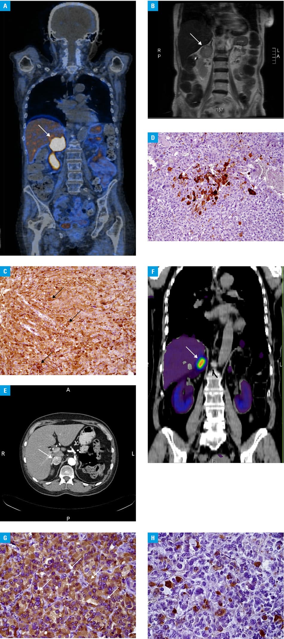
Patient 2 was a 61‑year‑old woman with a 2‑month history of weight gain, proximal myopathy, depressive disorders, abdominal pain, diabetes mellitus of recent onset, and worsening control of hypertension. Physical examination revealed facial and leg edema, plethoric face, dermal and muscle atrophy and moderate central obesity. Laboratory results are presented in Supplementary material, Table S1. Abdominal CT scan and SRI showed a 30‑mm mass in the right adrenal gland, with radiological suspicion of pheochromocytoma (Figure 1E and 1F). After pharmacological treatment of the patient, the tumor was removed laparoscopically without complications. Histological examination confirmed pheochromocytoma with focal expression of chromogranin A and ACTH‑secreting cells (Figure 1G and 1H). Currently, the patient is in complete remission and does not require any treatment.
Ectopic ACTH syndrome caused by pheochromocytoma is extremely rare, but should be considered as a possible source of ACTH production as it may be a life‑threatening condition if not diagnosed early and treated properly. A longer history of CS and cardiac or renal burden are associated with worse prognosis.
- Nijhoff MF, Dekkers OM, Vleming LJ, et al. ACTH‑producing pheochromocytoma: clinical considerations and concise review of the literature. Eur J Intern Med. 2009; 20: 682‑685. | Crossref
- Ilias I, Torpy DJ, Pacak K, et al. Cushing’s syndrome due to ectopic corticotropin secretion: twenty years’ experience at the National Institutes of Health. J Clin Endocrinol Metab. 2005; 90: 4955‑4962. | Crossref
- Lacroix A, Feelders RA, Stratakis CA, Nieman LK. Cushing’s syndrome. Lancet. 2015; 386: 913‑927. | Crossref
- Kubicka E, Zawadzka K, Syrycka J, et al. A case of gastrinoma associated with ectopic Cushing syndrome. Pol Arch Intern Med. 2020; 130: 328‑329. | Crossref
- Ballav C, Naziat A, Mihai R, et al. Mini‑review: pheochromocytomas causing the ectopic ACTH syndrome. Endocrine. 2012; 42: 69‑73. | Crossref
SUPPLEMENTARY MATERIAL
ARTICLE INFORMATION