Calcification as a cause of potential false‑positive findings on bone scintigraphy verified with 68Ga-PSMA-11 PET/CT: a case report


 CC BY-NC-SA 4.0
Calcification as a cause of potential false‑positive findings on bone scintigraphy verified with 68Ga-PSMA-11 PET/CT: a case report
Prostate cancer (PCa) is the second most common cause of cancer deaths among men in Poland. It is usually a slowly growing tumor, but sometimes it can metastasize early, most often to lymph nodes and bones.
Despite various imaging methods used in suspected cancer recurrence or bone metastases, such as scintigraphy, contrast enhanced computed tomography, or whole‑body magnetic resonance, positron emission tomography / computed tomography (PET / CT) targeted at the prostate‑specific membrane antigen (PSMA) expressed on PCa cells is the best solution for PCa imaging. It showed a higher detection rate than other imaging modalities for disease recurrence with detection rates of 33%, 45%, 59%, 75%, and 95% for prostate‑specific antigen (PSA) levels 0 to 0.19, 0.2 to 0.49, 0.5 to 0.99, 1 to 1.99, and ≥2 ng/ml, respectively.1,2
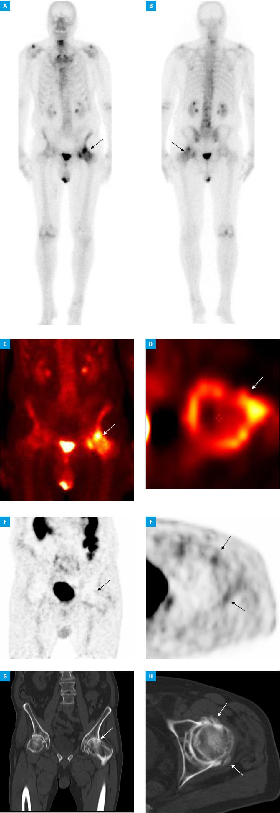
A 76‑year‑old man with prostate adenocarcinoma, the initial PSA level of 8.5 ng/ml, 1 year after robotic assisted laparoscopic prostatectomy with Gleason score 3 + 3 = 6, and with biochemical recurrence with the PSA level of 0.446 ng/ml (PSA doubling time of 3 months) underwent bone scintigraphy. It showed an intense diffuse “hot” lesion in the left femur (Figure 1A and 1B). Single‑photon emission computed tomography (SPECT) also revealed an intense uptake around the femoral head (Figure 1C and 1D). Due to an atypical uptake pattern for bone metastasis on the SPECT image and in order to exclude other metastatic lesions before planned radiotherapy, the patient was referred for Gallium 68 (68Ga) PSMA‑11 PET / CT. Surprisingly, it did not show any abnormal uptake. No uptake was seen near the bone on PET in the transverse section corresponding to SPECT. The CT image of the same plane showed degenerative changes in the left hip with calcifications in the surrounding soft tissue (Figure 1E–1H).
Bone scintigraphy is the first‑choice method in case of a suspicion of bone metastasis caused by PCa as well as other types of cancer. The increased uptake (”hot” lesions) on technetium 99m methylene diphosphonate scintigraphy (99mTc‑MDP) could be seen in all processes (benign and malignant) which activate osteoclasts. Our case was a false‑positive finding owing to the fact that the activated osteoblast in the calcification of soft tissue resembled a metastatic lesion in the whole‑body image. In the literature, there are a few cases describing unusual calcifications on 99mTc‑MDP, for example, in vascular disease, physical exertion, cold exposure, or specific lesions after liver transplantation.3,4 The majority of them are associated with hypercalcemia after kidney transplantation or calcification after bone fracture. Our patient had neither a history of bone trauma nor an elevated calcium level.
Based on the recent European Association of Urology recommendations, a patient with biochemical recurrence with PSA exceeding 0.2 ng/ml could be offered 68Ga‑PSMA‑11 PET / CT.5 According to the reimbursement rules in Poland, it could be offered only to patients with a suspicion of bone metastasis and uncertain findings on bone scintigraphy. The false‑positive findings on bone scintigraphy could have strong clinical implications for treatment recommendation. This case showed that nowadays, hybrid images combining functional and anatomical images are required in case of any unclear picture.
- Perera M, Papa N, Roberts M, et al. Gallium‑68 prostate‑specific membrane antigen positron emission tomography in advanced prostate cancer‑updated diagnostic utility, sensitivity, specificity, and distribution of prostate‑specific membrane antigen‑avid lesions: a systematic review and meta‑analysis. Eur Urol. 2020; 77: 403‑417. | Crossref
- Fanti S, Minozzi S, Antoch G, et al. Consensus on molecular imaging and theranostics in prostate cancer. Lancet Oncol. 2018; 19: e696‑e708. | Crossref
- Castaigne C, Martin P, Blocklet D. Lung, gastric, and soft tissue uptake of Tc‑99m MDP and Ga‑67 citrate associated with hypercalcemia. Clin Nucl Med. 2003; 28: 467‑471. | Crossref
- Caobelli F, Paghera B, Pizzocaro C, Guerra UP. Extraosseous myocardial uptake incidentally detected during bone scan: report of three cases and a systematic literature review of extraosseous uptake. Nucl Med Rev Cent East Eur. 2013; 16: 82‑87. | Crossref
- Compilation of all Guidelines. European Association of Urology. http://uroweb.org/guidelines/compilations‑of‑all‑guidelines/. Accessed December 12, 2020.
ARTICLE INFORMATION