Ecthyma gangrenosum as the primary manifestation of acute myeloid leukemia in a previously healthy patient


 CC BY-NC-SA 4.0
Ecthyma gangrenosum as the primary manifestation of acute myeloid leukemia in a previously healthy patient
Ecthyma gangrenosum (EG) is a rare cutaneous infection that typically manifests in immunocompromised individuals and may be often associated with Pseudomonas aeruginosa infection.
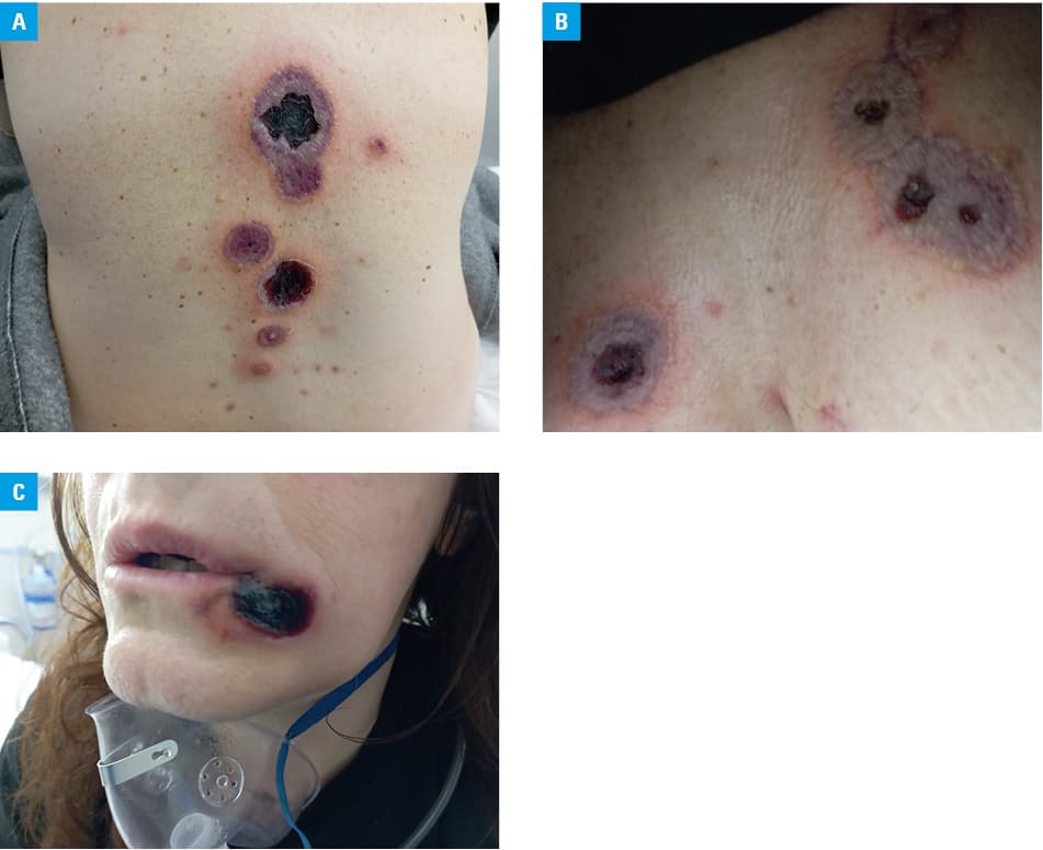
A 50‑year‑old, previously healthy woman reported to the emergency department due to deterioration of a rash that had started 14 days before and fever with an onset 4 days before. At the time of presentation, the skin lesions appeared as gangrenous ulcers with black scabs surrounded by a red halo, and were localized mainly on the trunk, chest wall, and labial commissures (Figure 1A–1C). The patient had undergone a laboratory workup 6 months prior, and the results were normal.
Following the onset of fever episodes and rash deterioration, a diagnostic workup was undertaken. Laboratory investigations revealed normocytic normochromic anemia (hemoglobin, 9.0 g/dl; reference range [RR], 12.0–16.0 g/dl; mean corpuscular volume, 81 fl; RR, 80–99 fl; reticulocytes, 0.23%; RR, 0.2%–2%), leukocytosis (white blood cells [WBCs], 31.2 K/μl; RR, 3.8–10.5 K/μl; neutrophils, 19.4%; RR, 45%–75%; lymphocytes, 13.1%; RR, 20%–51%; monocytes, 67.5% atypical cells; RR, 2.0%–11.0%), and thrombocytopenia (platelets, 20 Κ/μl; RR, 150–450 K/μl). Peripheral blood smear showed 67% of blast cells in the WBC count, and flow cytometry was indicative of acute monoblastic / monocytic leukemia (leukemic blasts revealed CD34+, CD33+, CD13+, CD64+, CD300E+, CD4+, T1T+, CD38+, CD11a+, CD11b+, CD11+/–, CD36+/–, CD16+/–, HLADR+/–, MPO+/–, and CD14+/–). Bone marrow aspirate examination confirmed the diagnosis of acute myeloid leukemia (AML), while molecular karyotyping did not reveal any specific chromosome abnormality. A whole‑body computed tomography scan was unremarkable. A skin biopsy was performed, and the histopathological examination of the lesion specimen revealed vascular necrosis with several surrounding bacteria. Gram‑stained sections showed gram‑negative rods surrounding the necrotic vessels, indicative of Pseudomonas aeruginosa infection; blood and lesion cultures confirmed the diagnosis. Following a decrease in the levels of hemoglobin and a sharp increase in inflammatory marker concentrations (C‑reactive protein, 41.8 mg/dl; RR, 0–0.8 mg/dl, procalcitonin, 32.9 ng/ml; RR, <0.2 ng/ml, erythrocyte sedimentation rate, 148 mm; RR, 0–20 mm, adjusted for age, lactate dehydrogenase, 888 U/l; RR, 135–214 U/l, β2‑microglobulin, 5.17 mg/l; RR, 1.42–3.21 mg/l), transfusion of red blood cells was performed, and antibiotic therapy with meropenem, linezolid, vancomycin, and isavuconazole was initiated. Despite fever deterioration, chemotherapy was introduced on day 5 of hospitalization, according to the standard schedule (idarabucin 12 mg/m2 for 3 days and aracytin 100 mg/m2 for 7 days). Meanwhile, the lesions evolved into necrotic ulcers involving the chest wall, oral cavity, and trunk. The patient developed sepsis and died within 3 weeks of admission to the hospital.
EG may initially present as an erythematous macule and may evolve into a hemorrhagic vesicle that turns into a gangrenous ulcer with necrotic eschar.1 Apart from immunocompromised patients, it can also affect otherwise healthy, immunocompetent individuals, with leukemia being one of the main predisposing conditions.2 Thus, EG occurrence in a previously healthy individual may signal undiagnosed immunodeficiency and warrants further investigation. Acute monocytic and monoblastic leukemias, occurring most commonly in young and adult people, respectively, account for less than 5% of all AML cases. Some individuals may present with bleeding disorders, whereas cutaneous involvement is common.2 Reports of AML cases presenting with EG as the primary manifestation are scant3; therefore, recognizing the underlying cause of EG is the key to a proper therapeutic approach.
- Vaiman M, Lazarovitch T, Heller L, Lotan G. Ecthyma gangrenosum and ecthyma‑like lesions: review article. Eur J Clin Microbiol Infect Dis. 2015; 34: 633‑639. | Crossref
- Biscaye S, Demonchy D, Afanetti M, et al. Ecthyma gangrenosum, a skin manifestation of Pseudomonas aeruginosa sepsis in a previously healthy child: a case report. Medicine (Baltimore). 2017; 96: e5507. | Crossref
- Hadano Y, Yoshida‑Sakai N, Imamura Y, et al. Acute myeloid leukaemia presenting with ecthyma gangrenosum as the first manifestation: a case report. Medicine (Baltimore). 2021; 100: e25867. | Crossref
ARTICLE INFORMATION