Bilateral spontaneous carotid artery dissection concomitant with early-stage COVID-19


 CC BY-NC-SA 4.0
Bilateral spontaneous carotid artery dissection concomitant with early-stage COVID-19
In December 2021, a previously healthy 50‑year‑old woman with no history of hypertension and smoking was admitted to the hospital with symptoms of acute ischemic stroke that had started 6 hours earlier. On admission, mild paresis of the upper left limb without sensory deficits was found, marked particularly in its distal part (National Institute of Health Stroke Scale [NIHSS] 1). She also reported dry cough, headache, and rhinitis lasting for 1 day. Her body temperature, blood pressure (114/75 mm Hg), and electrocardiography findings (sinus rhythm, 80 bpm) were normal. She denied any recent head or neck trauma, but reported previous hysterectomy and occasional migraine‑like headaches. Family history was negative for serious cardiovascular or connective tissue diseases.
A Doppler examination showed bilateral internal carotid artery (ICA) dissection (Figure 1A and 1B) and neuroimaging by computed tomography (CT) revealed a small acute ischemic lesion in the right parietal lobe.
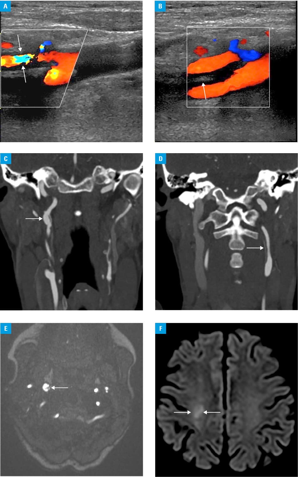
The peak C‑reactive protein concentration was 14.8 mg/l (upper limit of normal [ULN], 0.5 mg/l). Complete autoimmune screening was negative. The lipid profile results were as follows: total cholesterol, 140.5 mg/dl (ULN, 190 mg/dl); high‑density lipoprotein cholesterol, 56.6 mg/dl (normal range >45 mg/dl); low‑density lipoprotein cholesterol, 67.1 mg/dl (ULN, 100 mg/dl); triglycerides, 84.2 mg/dl (ULN, 150 mg/dl). A routine nasopharyngeal swab test performed on admission was positive for SARS‑CoV‑2. Subsequent CT angiography (CTA) documented dissection of both ICAs with intramural hematoma in the left ICA (Figure 1C and 1D).
Following a multidisciplinary discussion, conservative management was chosen. During the hospitalization, the patient’s neurological state improved—a reduction of the left upper limb paresis was observed. She was discharged home in a good condition (NIHSS 0).
Two months later, the patient was referred for a CTA examination, and a head‑to‑pelvis imaging study revealed neither fibromuscular dysplasia (as underlying arteriopathy) nor other arterial dissections.1 Magnetic resonance imaging showed partial remission of the vascular wall injury on both ICAs and revealed remnants of recent cerebral ischemia as documented by moderate hyperintensity in diffusion‑weighted imaging (Figure 1E and 1F). The results of 24‑hour ambulatory blood pressure monitoring were normal.
The final diagnosis was spontaneous bilateral ICA dissection with right‑sided ischemic stroke associated with an early stage of mild SARS‑CoV‑2 infection. Other predisposing factors may have included the winter season and migraine‑like headaches.2
An association of spontaneous artery dissection with COVID‑19 has been reported previously, including recent cases of dissections involving the coronary arteries and the aorta.3 Until recently, reports on dissections in other vascular beds have been scant. Our case confirms that in susceptible patients an ICA dissection also may be associated with SARS‑CoV‑2 infection, even at the early stage of mildly symptomatic COVID‑19.4
- Talarowska P, Dobrowolski P, Klisiewicz A, et al. High incidence and clinical characteristics of fibromuscular dysplasia in patients with spontaneous cervical artery dissection: the ARCADIA‑POL study. Vasc Med. 2019; 24: 112‑119. | Crossref
- Debette S, Leys D. Cervical‑artery dissections: predisposing factors, diagnosis, and outcome. Lancet Neurol. 2009; 8: 668‑678. | Crossref
- Cannata S, Birkinshaw A, Sadom D, et al. Spontaneous coronary artery dissection after COVID‑19 infection presenting with ST segment elevation. Eur Heart J. 2020; 41: 4602. | Crossref
- Januszewicz A, Wojciechowska W, Prejbisz A, et al. Impact of the COVID‑19 pandemic on blood pressure control and cardiovascular risk profile in patients with hypertension. Pol Arch Intern Med. 2021; 131: 16129. | Crossref
ARTICLE INFORMATION