Thrombotic microangiopathy in a patient with antiphospholipid syndrome and malignant hypertension

 CC BY 4.0
Thrombotic microangiopathy in a patient with antiphospholipid syndrome and malignant hypertension
Kidney involvement is a common finding in patients with systemic lupus erythematosus (SLE) and with antiphospholipid syndrome (APS). Malignant hypertension (MHT) is a rare but still present hypertensive emergency that influences cardiovascular risk.1 Kidney biopsy (KB) allows clinicians to diagnose the type of renal involvement.
A 35‑year‑old man was referred to a hospital due to elevated serum creatinine, proteinuria, anemia, and thrombocytopenia. Until the time of hospitalization the patient had not received any treatment for chronic diseases. For several days before the admission, the patient had reported dyspnea on exertion, abdominal pain, and nocturia. On admission his blood pressure was 200/125 mm Hg. Laboratory tests showed hemoglobin at 10.1 g/dl (reference range [RR], 13.7–17.5 g/dl), platelet count of 88 000/µl (RR, 132 000–370 000/µl], proteinuria of 1.77 g/24 hours (RR <0.15 g/24 hours), with microscopic hematuria, serum creatinine at 2.74 mg/dl (RR, 0.73–1.18 mg/dl), estimated glomerular filtration rate of 29 ml/min/1.73 m2, and lactate dehydrogenase of 257 U/l (RR, 120–246 U/l). There were no schistocytes on blood smear. Direct Coombs test was positive. Prolonged activated partial thromboplastin time (99.3 s; RR, 24–36 s) was not normalized by a correction test. The presence of lupus anticoagulant was confirmed using diluted Russell viper venom time. Immunological investigations revealed positive antinuclear antibodies (titer 1:320), positive immunoglobulin (Ig) G anticardiolipin and IgM anti-β2glycoprotein‑I antibodies, while anti–double‑stranded DNA antibodies were negative. The level of C3 was 0.8 g/l (RR, 0.79–1.52 g/l), and that of C4 0.15 g/l (RR, 0.16–0.38 g/l). ADAMTS‑13 activity was 78% (RR, 60%–121%).
The patient was screened for hypertension‑mediated organ damage. Echocardiography showed left ventricular and left atrial enlargement, left ventricular hypertrophy and reduced ejection fraction (40%), mitral valve insufficiency, and mild pericardial effusion. Fundoscopy revealed optic disc swelling with blurred disc margins, hemorrhages, and cotton wool exudates, which corresponded to grade IV of Keith–Wagener–Baker hypertensive retinopathy. Brain magnetic resonance imaging showed enlarged perivascular spaces, white matter hyperintensities, and a few vascular lesions. Ultrasound showed the right and left kidney longitudinal size of 105 mm and 98 mm, respectively (RR, 100–120 mm). To define the type of kidney involvement, KB was performed.
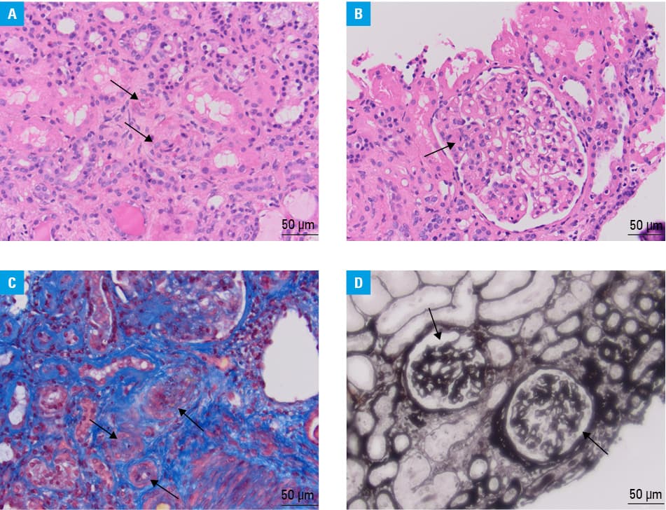
KB revealed acute thrombotic microangiopathy (TMA) with fibrin thrombi within the arterioles and fibrinoid necrosis of the arteriolar wall (Figure 1A), thrombi within segmentally dilated glomerular capillaries (Figure 1B), “onion skin” lesion with myointimal thickening and obliteration of the arteriolar lumen (Figure 1C), and ischemic wrinkling and collapse of the glomerular capillary wall (Figure 1D). Immunofluorescence staining showed no immune deposits.
As the clinical picture might have suggested SLE, APS, and MHT, KB seemed crucial for a differential diagnosis. KB revealed TMA and arteriolar wall fibrinoid necrosis.
The differential diagnosis of TMA should include SLE, APS, scleroderma, thrombotic thrombocytopenic purpura, infection‑related or complement‑mediated hemolytic uremic syndrome, drug‑induced TMA, cancer‑associated TMA, and MHT.2 The patient met the 2023 American College of Rheumatology / European League Against Rheumatism antiphospholipid syndrome classification criteria.3 Kidney involvement in SLE without immune deposits is very rare.4 MHT is a still present hypertensive emergency, with annual incidence around 1–2 cases per 100 000.5 Both APS and MHT are recognized causes of renal TMA, and both might be associated with pathogenic variants in complement genes.2 In the presented patient most, if not all, symptoms can be explained by APS and MHT, which makes SLE diagnosis unlikely.
- Mancia G, Kreutz R, Brunström M, et al. 2023 ESH Guidelines for the management of arterial hypertension. The Task Force for the management of arterial hypertension of the European Society of Hypertension Endorsed by the European Renal Association (ERA) and the International Society of Hypertension (ISH). J Hypertens. 2023; 41: 1874‑2071.
- Genest DS. Patriquin CJ, Licht C, et al. Renal thrombotic microangiopathy: a review. Am J Kidney Dis. 2023; 81: 591‑605. | Crossref
- Barbhaiya M, Zuliy S, Naden R, et al. 2023 ACR/EULAR antiphospholipid syndrome classification criteria. Arthritis Rheumatol. 2023; 75: 1687‑1702.
- Chen W, Liang S, Zuo K, et al. Clinicopathological features and outcomes of SLE patients with renal injury characterised by thrombotic microangiopathy. Clin Rheum. 2021; 40: 2735‑2743. | Crossref
- Shantsila A, Lip GY. Malignant hypertension revisited ‑ does this still exist? Am J Hypertens. 2017; 30: 543‑549. | Crossref
ARTICLE INFORMATION