Giant rhinophyma in a heart and kidney transplant recipient: giant oaks from little acorns grow


 CC BY-NC-SA 4.0
Giant rhinophyma in a heart and kidney transplant recipient: giant oaks from little acorns grow
Organ transplant recipients are prone to skin complications, such as neoplasms and infections.1 Rhinophyma is a nasal deformity characterized by an excessive growth of sebaceous glands and the underlying connective tissue, resulting in disfigurement. Rosacea is a precursor condition leading to the onset of rhinophyma. Cases of giant rhinophyma lesions are infrequently observed.
We present a case of a white man who underwent heart transplantation in 1998 at the age of 50 due to ischemic cardiomyopathy. In 2006, he received a subsequent renal transplant from a deceased donor due to calcineurin inhibitor toxicity. His immunosuppressive medications included mycophenolate mofetil, cyclosporine (CyA), and prednisone. Before kidney transplant, he had no known skin issues.
About 4 years after the renal transplant, the patient noticed a flushing and later redness on the skin of his nose. Initially, the observed cutaneous alterations were suspected to be drug‑induced. The patient attributed them to a transition from modified cyclosporine to its generic equivalent. However, reinitiation of the original medication failed to demonstrate improvement or arrest progression of the changes. In December 2013, a definitive diagnosis of rosacea was established. Topical therapy with metronidazole and benzoyl peroxide along with oral tetracycline proved ineffective. The patient experienced progressive skin thickening and formation of nodules in the nasal region, findings highly suggestive of a diagnosis of rhinophyma. The mass growth accelerated in 2017. The patient’s medical history did not include alcohol abuse. His dermatologic history included herpes zoster of the trunk skin with sequelae of postherpetic neuralgia and scarring (2011) and multiple squamous cell carcinomas excised from the dorsal aspects of both hands (2016). The patient’s skin condition led to embarrassment, prompting avoidance of social interactions, and causing severe depression.
After the transplantation, the patient attended regular appointments at the transplant clinic. He was reluctant to undergo cosmetic surgery due to anticipated wound healing complications on immunosupressive treatment. After extensive discussions to clarify the benefits and potential risks of the proposed surgical procedure, he ultimately consented to it at the age of 71. At this point, physical examination revealed a large, swollen, irregular mass on the tip, dorsum, and both alae of the nose, causing partial nostril obstruction and breathing difficulties (Figure 1A–1C). The other regions of the head and neck showed no abnormalities. On histopathologic examination, focal thinning of the epidermis was observed. Additionally, sebaceous gland hyperplasia with accompanying lymphocytic infiltration and interstitial fibrosis were noted, along with mild interstitial edema consistent with the diagnosis of rhinophyma.2
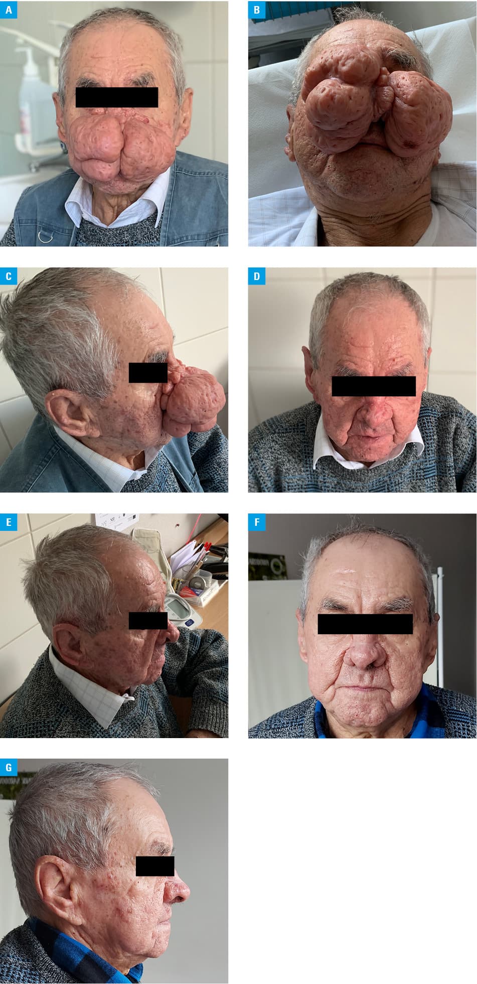
Subsequently, the patient was referred to the Department of Plastic and Reconstructive Surgery and underwent rhinophyma reduction surgery in November 2019. The cosmetic outcome was considered satisfactory by the patient (Figure 1D and 1E), leading to a resumption of social interactions. Throughout the 4‑year follow‑up, the patient remained free of recurrence (Figure 1F and 1G).
Pathophysiology of rhinophyma remains poorly understood. Vascular abnormalities contribute to a local production of transforming growth factor β1 (TGF-β1), with a potential to induce fibrosis and consequent cutaneous thickening.3 Of particular interest, calcineurin inhibitors, such as CyA and tacrolimus have been shown to induce TGF-β1 receptor–triggered signaling cascades.4 This suggests a possible link between cyclosporine use by our patient and rhinophyma development.
In conclusion, we report a case of giant rhinophyma, characterized by an esthetically displeasing appearance of the nose and facial features. In the past 2 decades, less than 25 cases of giant rhinophyma have been documented in the literature.5 Medical interventions have typically yielded unsatisfactory outcomes, emphasizing the necessity for rhinophyma reduction surgery in such cases.
- Thet Z, Lam AK, Pham T, et al. Clinical and economic burden of benign and malignant skin lesions in renal transplant recipients. Intern Med J. 2023; 53: 2042‑2049. | Crossref
- Schüürmann M, Wetzig T, Wickenhauser C, et al. Histopathology of rhinophyma ‑ a clinical‑histopathologic correlation. J Cutan Pathol. 2015; 42: 527‑535. | Crossref
- Cribier B. Pathophysiology of rosacea: redness, telangiectasia, and rosacea. Ann Dermatol Venereol. 2011; 138: S184‑S191. | Crossref
- Akool ES, Doller A, Babelova A, et al. Molecular mechanisms of TGF beta receptor‑triggered signaling cascades rapidly induced by the calcineurin inhibitors cyclosporin A and FK506. J Immunol. 2008; 181: 2831‑2845. | Crossref
- Cabrini G, La Torre P, Buonamico A, et al. A case report of giant rhinophyma. Otorhinolaryngol Head Neck Surg. 2019; 4: 1‑3. | Crossref
ARTICLE INFORMATION