


 CC BY 4.0
Adrenal oncocytic neoplasms are rare entities, with 289 cases described in the literature to date, including our case.1 These tumors are usually diagnosed incidentally, are not hormonally active, and predominantly affect the left adrenal gland. The median age at diagnosis is 44 years, with predominance in women.1,2 Adrenal oncocytic tumors are classified into 3 categories: oncocytoma, adrenal oncocytic neoplasm of uncertain malignant potential (AONUMP), and the oncocytic variant of adrenocortical carcinoma.3
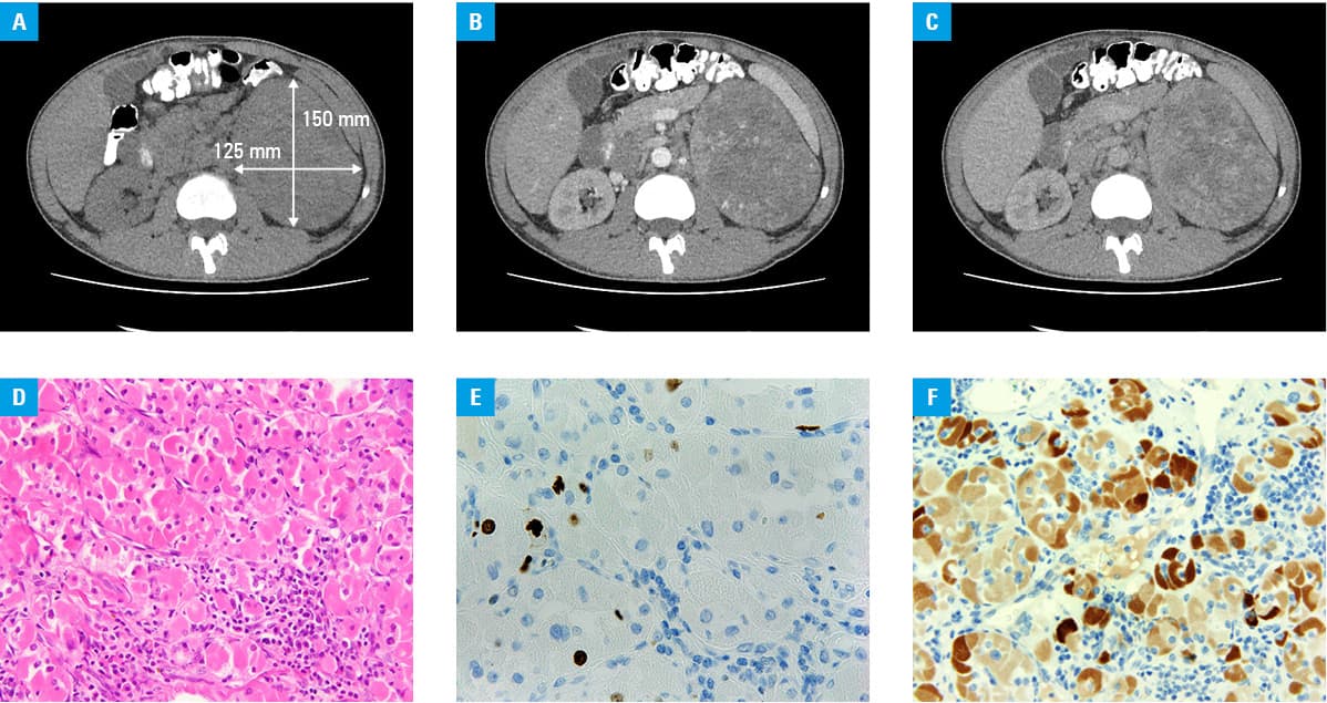
A 30‑year‑old man was admitted to a hospital after an incidental finding of a large left adrenal gland mass on ultrasound examination performed for newly diagnosed hypertension (180/120 mm Hg), and concomitant hypokalemia (2.85 mmol/l; reference range [RR], 3.5–5.1 mmol/l). Subsequent computed tomography (CT) showed a solid, heterogeneous tumor, highly suspicious of malignancy, measuring 150 mm × 125 mm × 100 mm. The lesion exhibited necrosis, calcification, and significant contrast enhancement in the solid components. Displacement of adjacent organs and a blurred border between the lesion and the left kidney were observed, but there were no obvious signs of infiltration (Figure 1A–1C).
Biochemical assays excluded pheochromocytoma and endogenous hypercortisolism. Serum concentrations of dehydroepiandrosterone sulfate, 17‑hydroxyprogesterone, estradiol, and androstenedione were within normal ranges. Adrenocorticotropic hormone (ACTH) levels were elevated to 450.3 pg/ml (RR, 6–56 pg/ml). The aldosterone‑to‑renin ratio was 10.35 (RR <20–40), with a plasma aldosterone concentration of 20.7 pg/ml (RR, 10–160 pg/ml) and low plasma renin activity (PRA, 0.2 ng/ml/h; RR, 0.51–2.64 ng/ml/h). The evaluation of aldosterone and PRA was performed at a serum potassium level of 3.39 mmol/l, as normokalemia (3.5–5.1 mmol/l) could not be achieved, and finally Conn syndrome was diagnosed.
The patient underwent laparoscopic adrenalectomy. Histopathological analysis (and subsequent re‑examination) identified the tumor as AONUMP. The lesion showed the absence of major malignant features (0–1 mitoses per 50 high‑power fields, no atypical mitoses, and no venous invasion; Figure 1D) and only 1 minor malignant feature (tumor size >10 cm). Degenerative changes, including necrosis and hemorrhage, were present, but no capsular invasion was observed. The Ki‑67 proliferation index was 4.5% (Figure 1E). The tumor cells were strongly positive for Melan‑A (Figure 1F), weakly positive for synaptophysin and α-inhibin, and negative for chromogranin A.
Postoperatively, the patient’s blood pressure and serum potassium level normalized. Serum ACTH level decreased to 205.7 pg/ml, and adrenal insufficiency was excluded based on adequate morning serum cortisol levels. Follow‑up CT scans at 2 and 8 months after surgery showed no signs of recurrence.
Oncocytic adrenal tumors, which often mimic adrenal carcinoma on CT, generally have a favorable prognosis, as compared with the latter. The 5‑year survival rate is 100% for oncocytomas, 88% for AONUMP, and 47% for the oncocytic variant of adrenocortical carcinoma. For stage III and IV adrenocortical carcinoma, survival rates drop to 24% and 0%, respectively.2,4 Laparoscopic surgery is usually curative for AONUMP, and chemotherapy is typically not required.2-4 However, due to a risk of local or distant recurrence, close follow‑up is essential.
Even in low‑risk cases, follow‑up protocols should include CT scans every 3 months for 4–5 years.2,4 Hormonal activity of adrenal oncocytomas, though rare, typically resolves after the tumor removal.
This case highlights the limitations of imaging in assessing suspicious adrenal masses and emphasizes the importance of comprehensive biochemical and histopathological examinations in determining prognosis and follow‑up duration.
- Coppola Bottazzi E, Gambardella C, Mongardini FM, et al. Prognosis of adrenal oncocytic neoplasms (AONs): literature review of 287 cases and presentation of the oldest patient. J Clin Med. 2023; 12: 6925. | Crossref
- Chin CP, Grauer R, Ucpinar B, et al. Oncocytic adrenocortical neoplasm of borderline uncertain malignant potential diagnosed after robot‑assisted adrenalectomy case report. BMC Urol. 2023; 23: 60. | Crossref
- Ramamoorthi S, Jindal B, Deivasigamani A, Srinivas BH. Oncocytic adrenal cortical neoplasm of uncertain malignant potential ‑ a rare case report. Indian J Pathol Microbiol. 2022; 65: 692‑694. | Crossref
- Kanitra JJ, Hardaway JC, Soleimani T, et al. Adrenocortical oncocytic neoplasm: a systematic review. Surgery. 2018; 164: 1351‑1359. | Crossref
ARTICLE INFORMATION