


 CC BY-NC-SA 4.0
The absence of coronary artery calcification has a high negative predictive value for obstructive coronary artery disease (CAD), even in patients with chest pain.1,2 However, there are exceptions to this rule. Here, we present a case of a 47‑year‑old man with hypertension, hypercholesterolemia, and a history of spinal surgery for scoliosis, who sought medical attention for moderate3 exertional dyspnea. Electrocardiography (Figure 1A) and echocardiography were unremarkable. Given the patient’s low risk factor–weighted clinical probability of CAD, coronary computed tomography angiography (CCTA) was scheduled.3
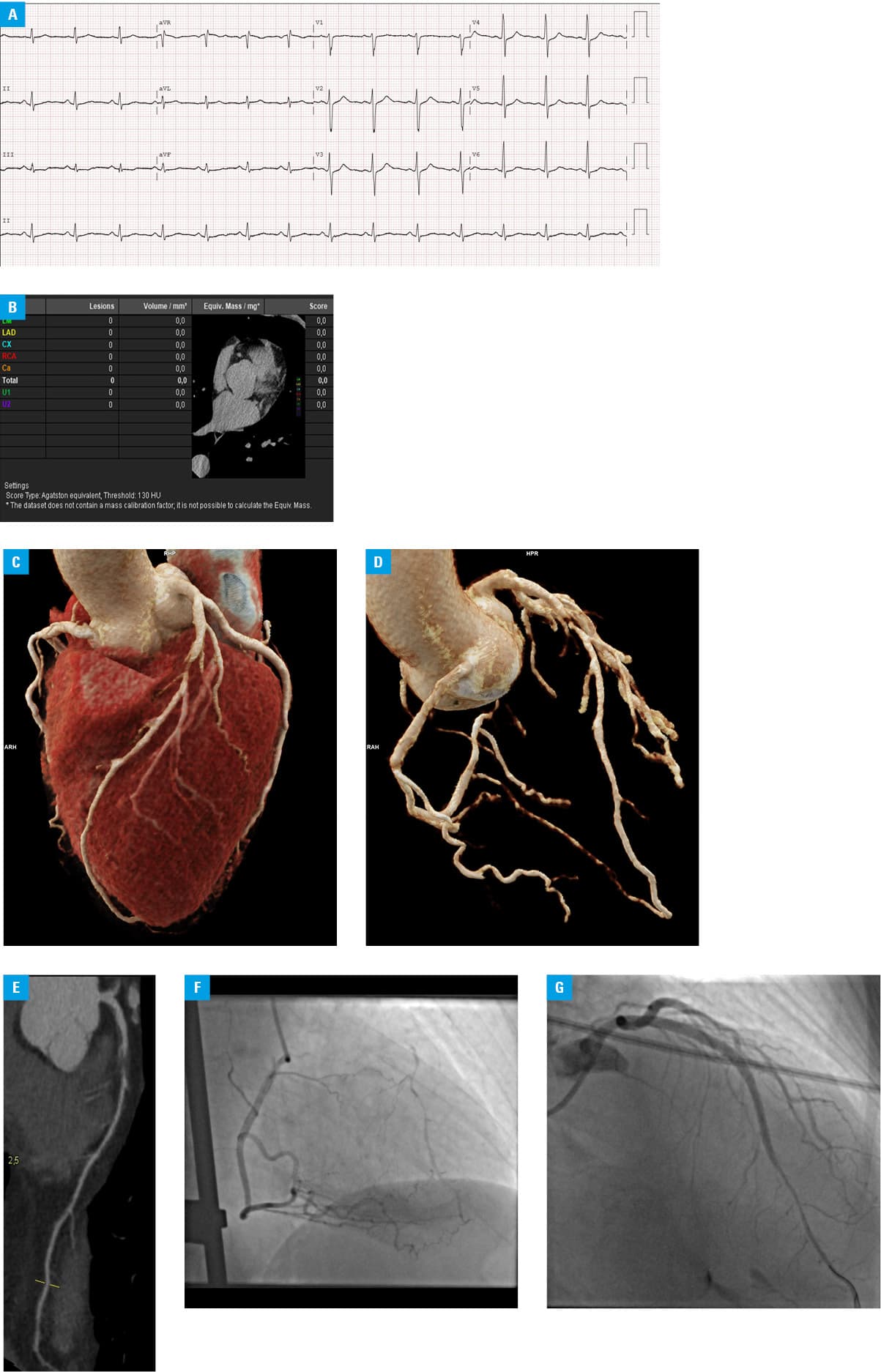
Abbreviations: Ca, calcified lesion (coronary artery); CX, left circumflex artery; HU, Houndsfield unit; LAD, left anterior descending artery; LM, left main coronary artery; RCA, right coronary artery; U1, user‑defined lesion type 1; U2, user‑defined lesion type 2
Despite the coronary artery calcium score (CACS) of 0, further examination showed a complete contrast loss in the left anterior descending artery (LAD; Figure 1B–1E). There were no abnormalities in the remaining coronary arteries. Coronary angiography confirmed complete LAD occlusion with good collateral circulation (Figure 1F). Percutaneous coronary intervention with stent implantation was performed (Figure 1G).
CACS evaluation is recommended when the pretest probability of CAD is 5%–15%, as it helps identify individuals with a CAD likelihood below 5%.3 In our patient, relying solely on a CACS‑based diagnosis might have led to a deferral of further testing. However, in a cohort study of symptomatic patients with obstructive CAD, a CACS score of 0 was most common in younger individuals, and its prevalence decreased progressively with patient age.4 In symptomatic patients, it is necessary to adopt a tailored diagnostic approach that considers risk factors, clinical presentation, and limitations of CACS in detecting noncalcified plaque before choosing between CACS and CCTA.
- Agha AM, Pacor J, Grandhi GR, et al. The prognostic value of CAC zero among individuals presenting with chest pain: a meta‑analysis. JACC Cardiovasc Imaging. 2022; 15: 1745‑1757. | Crossref
- Hecht HS. Coronary artery calcium scanning: past, present, and future. JACC Cardiovasc Imaging. 2015; 8: 579‑596. | Crossref
- Vrints C, Andreotti F, Koskinas KC, et al. 2024 ESC Guidelines for the management of chronic coronary syndromes: developed by the task force for the management of chronic coronary syndromes of the European Society of Cardiology (ESC) Endorsed by the European Association for Cardio‑Thoracic Surgery (EACTS). Eur Heart J. 2024; 45: 3415‑3537.
- Mortensen MB, Gaur S, Frimmer A, et al. Association of age with the diagnostic value of coronary artery calcium score for ruling out coronary stenosis in symptomatic patients. JAMA Cardiol. 2022; 7: 36‑44. | Crossref
ARTICLE INFORMATION