
How to manage adrenal incidentaloma in a hypertensive patient?
Key words: adrenal incidentaloma, adrenal tumor, pheochromocytoma, primary aldosteronism, secondary hypertension

 CC BY 4.0
How to manage adrenal incidentaloma in a hypertensive patient?
CC BY 4.0
Adrenal incidentaloma (AI) is a mass discovered on imaging not performed for suspected adrenal diseases. Technological advancements and increasing availability of imaging modalities, along with a growing awareness of preventive care and an aging population, have led to an increase in AI detection, generating diagnostic and therapeutic dilemmas for clinicians. Every patient with AI should undergo a careful evaluation to establish the etiology of the tumor and detect all hormonally active lesions, including those that cause secondary hypertension. Hormonal hypertension carries a higher risk of cardiovascular complications and target organ damage than essential hypertension, independently of blood pressure control. In this review, we discuss the current 2023 European Society of Endocrinology guidelines on the management of AIs in the context of other expert documents (including recommendations of the Polish Society of Endocrinology), with the main focus on 2 secondary forms of hypertension—primary aldosteronism and pheochromocytoma—that should not be missed in clinical practice.
What is adrenal incidentaloma?
The term “incidentaloma” was introduced in 1982 by Geelhoed and Druy,1 who recognized that with the advent of improved resolution in radiological techniques, clinicians were faced with the unfamiliar dilemma of early diagnosis of an asymptomatic adrenal tumor.
By definition,2 adrenal incidentaloma (AI) is a mass or masses discovered on imaging that was, in fact, performed for different reasons than the suspicion of any adrenal disease. This excludes, firstly, all cases when imaging is the consequence of the diagnostic process in patients with signs and symptoms of pheochromocytoma, hypercortisolism, hyperaldosteronism, hyperandrogenism, or other adrenal pathology (including adrenal insufficiency). Secondly, the discovery of an adrenal mass as a result of screening imaging in patients with a hereditary syndrome associated with an increased likelihood of developing adrenal tumors also does not meet the criteria for AI recognition. The last scenario that falls outside the strict definition of AI is when abdominal computed tomography (CT) is performed in patients with extra‑adrenal tumors—for staging or for follow‑up. A common clinical situation that leads to the discovery of AI is when abdominal CT is performed for symptoms such as abdominal or back pain, or gastrointestinal bleeding. AI can also be found on chest CT performed because of a suspicion of lung diseases (CT screening in smokers or exclusion of pulmonary embolism).
The role of ultrasonography in the diagnosis of AI is limited due to low resolution, reduced penetration, and a lack of standardization in the interpretation of results. Abdominal ultrasound can lead to a suspicion of AI; its presence should, however, be confirmed on other diagnostic modalities (with abdominal CT being the first choice in most patients, according to guidelines).2,3
Adrenal incidentaloma: workup and key issues
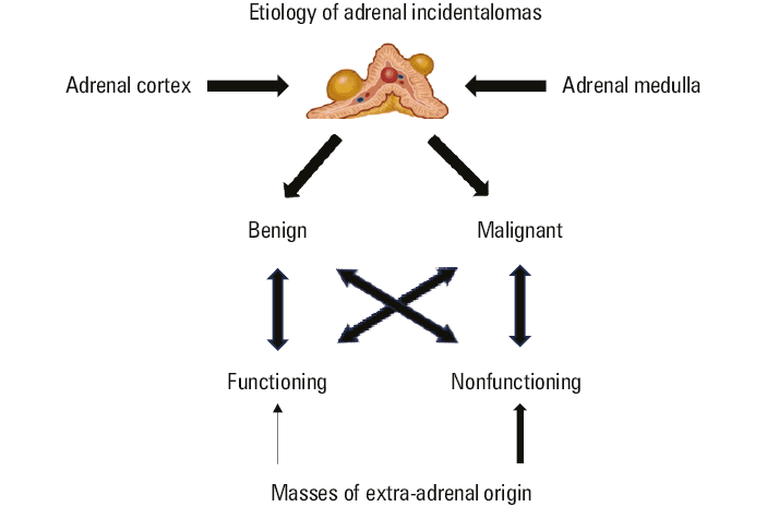
There are some fundamental questions to be asked during the workup of AI, and usually they are being answered in parallel. Adrenal masses originate either from the adrenal glands, cortex, or medulla, or from extra‑adrenal tissue (Figure 1).
Imaging is the most important tool to clarify the nature of the tumor; however, hormonal testing can also be highly useful or even critical. The most recent 2023 European Society of Endocrinology (ESE) clinical practice guidelines on the management of AIs,2 as well as the 2016 Polish Society of Endocrinology incidentaloma guidelines and the 2024 Polish Society of Endocrinology guidelines on the management of adrenocortical carcinoma,3,4 suggest similar diagnostic pathways in patients with adrenal tumors (Figure 2). They all strongly recommend to establish with the highest possible certainty if an adrenal mass is benign or malignant at the time of initial detection. In homogenous lesions with noncontrast CT enhancement lower than or equal to 10 Hounsfield units (HU), the risk of malignancy is virtually absent, and these tumors do not require further investigation to confirm their benign character.
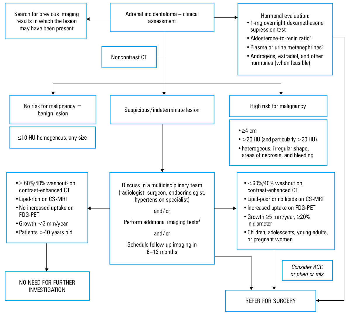
a In patients with concomitant hypertension or hypokalemia
b In patients with an adrenal lesion with a density >10 HU
c 60%/40% stands for absolute and relative contrast washout after 15 minutes3,4
d One of 3 modalities: CS‑MRI, contrast‑enhanced CT and / or FDG‑PET
Abbreviations: ACC, adrenocortical carcinoma; CS‑MRI, chemical shift–magnetic resonance imaging; CT, computed tomography; FDG‑PET, 18F‑fluorodeoxyglucose positron emission tomography; HU, Hounsfield unit; mts, metastases; pheo, pheochromocytoma
Benign tumors (eg, myelolipoma) are resected only if they are very large and cause a mass effect. In patients with homogenous lesions with a tumoral density between 10 and 20 HU and a diameter of less than 4 cm, other imaging modalities may be useful, or follow‑up imaging should be scheduled. In such cases, options should be individually discussed by a multidisciplinary team. If an adrenal mass is larger than or equal to 4 cm and heterogeneous, or has an unenhanced density above 20 HU (particularly >30 HU), the risk of malignancy is high. A multidisciplinary team should discuss proceeding to immediate surgery, which is the management of choice for most patients. As adrenocortical carcinoma (ACC) is an aggressive malignancy with poor prognosis,5 it is crucial to diagnose all cases of this tumor, which may be difficult.6 However, it has been widely recognized that most AIs display a benign behavior over time, and adrenalectomy should only be performed in a small proportion of patients, either because of a suspicious phenotype at the initial assessment or due to a significant growth at follow‑up. Even in patients with a “nonadenoma” phenotype of the tumor, the risk of malignancy on the postoperative histopathologic report is relatively low.7
After excluding malignancy, the second goal is to detect all hormonally active lesions.3 Despite the fact that most AIs are nonfunctioning, almost every patient diagnosed with AI should undergo a careful biochemical workup to rule out adrenal hormone excess that can be associated with endocrine hypertension.8 Only in frail patients with limited life expectancy may particular tests (such as the 1‑mg overnight dexamethasone test) be omitted.2 In some situations, the initial hormonal evaluation is inconclusive. Repeated hormonal testing—the “wait and retest” approach—can be useful, as functional incidentaloma is a progressive disease, and the presence of hormone excess often becomes more evident with time.9
It is worth stressing that the current guidelines2,3 limit the definition of AI and workup recommendations to lesions with a diameter greater than or equal to 1 cm. Patients with bilateral or multiple adrenal masses should undergo a clinical and hormonal assessment identical to that performed in patients with unilateral AI. Each adrenal lesion should be assessed individually at the time of initial detection, according to the same imaging protocol as for unilateral adrenal masses, to establish its phenotype.
The subgroups of patients that would benefit from an urgent assessment of an adrenal mass because of a higher likelihood of malignancy and clinically significant hormone excess are pregnant women and individuals below 40 years of age. If dedicated adrenal imaging is required, the use of magnetic resonance (MRI) rather than CT is preferred in children, adolescents, and pregnant women. Surgical resection rather than follow‑up is suggested if an adrenal mass is indeterminate on imaging in children, adolescents, pregnant women, and adults younger than 40 years.
Prevalence of adrenal incidentalomas in the general population
The prevalence of adrenal tumors in the general population is difficult to establish and can only be extrapolated from unselected imaging or autopsy studies. It varies depending on the source of data and patient selection (from general or reference centers). The rates of detection of adrenal tumors on radiological examinations have significantly increased in recent years. Several factors underlie this phenomenon, including technological advancements in imaging modalities, a growing awareness of preventive care, the rising number of diagnostic imaging centers, and an increasing prevalence of chronic disease, driven in large part by an aging population.
Ebbehoj et al10 performed a retrospective population‑based cohort study in the United States and reported an increase in the incidence rates of adrenal tumors (mainly incidentalomas) from 4.4 per 100 000 person‑years in 1995 to 47.8 in 2017, representing a more than 10‑fold increase. The study also revealed lower rates of malignancy, pheochromocytomas, and overt steroid hormone excess, as compared with previous reports.
Similarly, in a retrospective study conducted between 1997 and 2016 in China,11 the chronological distribution of patients also showed that the number of cases gradually increased each year. The authors of a more recent study from China,12 which included more than 25 000 healthy volunteers, reported that the prevalence of AIs also increased with age, ranging from 0.2% in the participants aged 18 to 25 years to 3.2% in those older than 65 years (with an overall prevalence of 1.4%). Other radiological series reported an even higher rate of detection in the oldest participants, from around 3% in adults older than 50 years and up to 10% in the patients older than 80 years.2
In autopsy series on large cohorts of patients, the reported prevalence of AI ranged from 1.05% to 8.7%, suggesting an overall prevalence of around 2%, increasing with age.2,13 In earlier estimates, the prevalence of clinically inapparent adrenal masses detected on autopsy was even lower than 1% for patients younger than 30 years and increased to 7% in patients aged 70 years or older; many of the lesions detected on autopsy were very small.14
Etiology of adrenal incidentalomas and the prevalence of functioning tumors
Over the past 3 decades, several studies11,15-20 have tried to establish the etiology of AIs and the prevalence of secreting tumors. According to the ESE data,2 based on most studies, 80%–85% of AIs are benign, unilateral or bilateral adrenocortical tumors (adrenal adenoma or bilateral adrenal hyperplasia). Other benign masses (3%–6%) that can be found in adrenals include myelolipomas, cysts and pseudocysts, ganglioneuromas, and schwannomas. Pheochromocytomas (either benign or malignant) constitute 1%–5% of all cases. Other possible malignancies are either ACCs (0.4%–4%) or metastases (3%–7%). Most of the adrenocortical tumors are nonfunctioning (50%–70%). Keeping in mind the heterogeneity of the study results, mild autonomous cortisol secretion seems to be the most common finding among secreting tumors (20%–50%), with primary aldosteronism being diagnosed in 2%–5% of cases and overt Cushing syndrome in 1%–4%.
Wide ranges of reported prevalence are being attributed both to the changing context of the studies and inclusion criteria, as well as to differences in the screening and diagnostic strategies. In a recent systematic review and meta‑analysis of the prevalence of functioning AIs,8 the overall proportion of secreting tumors was 27.5%. The final analysis included a total of 36 full‑text articles and more than 16 000 participants. The proportion of patients with hypertension was 53.7%. The prevalence of autonomous cortisol secretion, primary aldosteronism, pheochromocytoma, and even overt Cushing syndrome (contradicting the definition of AI) was assessed, yielding proportions of 11.7%, 4.4%, 3.8%, and 3.1%, respectively.
The findings of this meta‑analysis8 are contrary to the results of the abovementioned study by Ebbehoj et al10 on healthy volunteers, which reported an overall lower prevalence of functioning tumors (mean, 4.1%), reflecting presumably the lower rate of diagnostic workup in a real‑life context.
Should we screen individuals with adrenal incidentaloma for primary aldosteronism?
The ESE guidelines point to aldosterone‑to‑renin ratio (ARR)—the screening tool for primary aldosteronism (PA)—as one of the main biochemical tests in patients with AI to use in individuals with concomitant hypertension or unexplained hypokalemia.
The Endocrine Society and European Society of Hypertension21,22 both recommend broad screening for PA among patients with hypertension, with at least half of them being evaluated for this secondary form of hypertension. The selection of patients should be based on their clinical or biochemical features.
The subgroups of patients characterized by a higher prevalence of PA are therapy‑resistant individuals,23 those with severe hypertension (in theory conflicting with the definition of AI) or a family history of PA or early stroke (often unrecognized and also conflicting with the definition), young adults with hypertension, patients with hypertension and concomitant atrial fibrillation, and individuals with hypertension with hypokalemia or AI.
In many patients, screening with ARR is reliable even during an ongoing antihypertensive therapy without a problematic drug washout procedure.24,25 Literature data provide evidence that patients with PA, similarly to those with pheochromocytoma, have more pronounced target organ damage and cardiovascular complications disproportionate to blood pressure (BP) and the duration of hypertension. In a study by Savard et al,26 the prevalence of electrocardiographic and echocardiographic left ventricular hypertrophy (LVH) was about twice as high in the patients with PA as in matched controls with essential hypertension.
PA patients also had a significantly higher prevalence of coronary artery disease (adjusted odds ratio [OR], 1.9), nonfatal myocardial infarction (adjusted OR, 2.6), heart failure (adjusted OR, 2.9), and atrial fibrillation (adjusted OR, 5). Prolonged exposure to elevated aldosterone concentrations is associated with cardiac hypertrophy and fibrosis as well as numerous deleterious changes in the vessels: deterioration of endothelial function, intensification of oxidative stress, increase in arterial stiffness, decrease of vascular compliance, and finally atherosclerosis and resistant hypertension.27 Aldosterone‑mediated cardiac damage is not limited to LVH, but also includes impaired diastolic function and subclinical systolic dysfunction on echocardiography, as well as nonischemic fibrosis reflected by the presence of late gadolinium enhancement on MRI.28 In the kidney, the excess of aldosterone causes sodium retention and hyperfiltration, which leads to albuminuria and, finally, impaired renal function.
Therefore, the aim of treatment in PA should not be confined to BP normalization and hypokalemia correction, but should focus on restoring the water‑sodium balance on a hormonal level (including unblocking renin secretion)29. Current evidence convincingly demonstrates that both surgical and medical treatment strategies beneficially affect cardiovascular outcomes and mortality in the long term. This further supports the need for heightened vigilance on PA in a given individual and possibly for broadening the scope of screening at the population level.
Prevalence of primary aldosteronism in patients presenting with adrenal incidentaloma
PA may manifest across a wide spectrum of clinical and biochemical phenotypes, ranging from mild to overt.30 In a meta‑analysis by Sconfienza et al,8 the prevalence of PA in AI patients was 4.4%. In a study by Monticone et al,31 out of a total of 1672 primary care patients with hypertension, 5.9% were diagnosed with PA. The overall prevalence of PA increased with the severity of hypertension, from 3.9% in stage 1 hypertension to 11.8% in stage 3 hypertension. However, emerging evidence suggests that the prevalence of PA is much greater than previously recognized, and that nonclassic forms of renin‑independent aldosterone secretion may be common. “Nonclassic” or “subclinical” PA means that these patients do not meet the recommended thresholds of what is currently considered biochemical confirmation of PA but have subtle biochemical evidence of renin‑independent aldosteronism (renin suppression with inappropriately “normal” or high aldosterone levels). They can be mildly hypertensive or normotensive, but with a higher risk of developing hypertension.30 In young and otherwise healthy individuals, mostly women, spontaneous low baseline BP prevents the development of hypertension, despite aldosterone overproduction.32
Thus, some authors suggest screening not only hypertensive, but also normotensive patients with hypokalemia and / or AI, as well as those with borderline elevated BP, and patients who had very low BP levels in the past, with a substantial recent increase (but still in normotension ranges).33 During the diagnostic workup, the decisions on cutoffs separating essential hypertension from PA are in fact more or less arbitrary. Comparing with other hormones, such as cortisol, it is not surprising that subclinical aldosterone excess exists. The secretion of both hormones, aldosterone and cortisol, is present in 5%–21% of patients with PA.34 However, studies on steroid metabolome revealed an even higher percentage of glucocorticoid excess of up to 29%35 in patients with PA. In a study by Adolf et al,36 cortisol excess appeared to have an additional impact on cardiac remodeling in patients with PA, and treatment with either adrenalectomy or mineralocorticoid receptor antagonist had a positive effect on these parameters, especially in the individuals who manifested greater cortisol excess.
Diagnostic flowchart for primary aldosteronism patients presenting with adrenal incidentaloma
Current guidelines21,22 recommend that patients with a positive ARR undergo at least 1 test to definitively confirm or exclude the diagnosis of PA. However, in the setting of spontaneous hypokalemia below 3.5 mmol/l, low plasma renin levels, and a plasma aldosterone concentration above 20 ng/dl, confirmatory testing may be omitted.
When surgical treatment is feasible and desired by the patient, adrenal venous sampling (AVS) is a gold standard procedure to make the distinction between unilateral and bilateral adrenal disease.
Although noncontrast CT is sufficient to establish the diagnosis of adenoma in most patients with PA, it is convenient to perform the evaluation of abdominal vessels on contrast‑enhanced CT before AVS. Younger patients (<35 years) with spontaneous hypokalemia, marked aldosterone excess above 30 ng/dl, and unilateral adrenal lesions greater than 10 mm with radiological features consistent with cortical adenoma (and a normal contralateral adrenal gland), may not need AVS before proceeding to unilateral adrenalectomy. However, some experts recommend subtyping with AVS without exceptions.
Should we screen individuals with adrenal incidentaloma for pheochromocytoma?
The 2023 ESE guidelines2 recommend that every patient with AI should undergo careful clinical assessment for signs and symptoms of adrenal hormone excess and hormonal evaluation, even in asymptomatic cases. The presence of pheochromocytoma can be assessed by measuring plasma free or urine fractionated metanephrines. Hypertension, even paroxysmal, is not a sine qua non condition for screening for pheochromocytoma, as it is not more frequent in AI patients with pheochromocytoma that those without.37,38 Pheochromocytoma should be excluded if a tumor’s phenotype is not typical of a benign lesion (>10 HU). If adrenalectomy is considered, a differential diagnosis with pheochromocytoma is essential, because the latter requires specific presurgical and surgical management, and poses a risk of perioperative complications.4
Patients with pheochromocytoma display higher rates of cardiovascular events than those with essential hypertension, which cannot be attributed to differences in BP or other cardiovascular risk factors. In a retrospective case‑control study by Stolk et al,39 109 pheochromocytoma patients were assessed for cardiovascular events (ischemic heart disease, cerebrovascular accidents, and transient ischemic attacks) within 5 years prior to the diagnosis. These patients were matched to controls with essential hypertension. A significantly higher rate of patients with pheochromocytoma was shown to experience a cardiovascular event, as compared with the hypertensive patients (13.8% vs 1.1%; OR, 14.4), despite the fact that the BP level was lower in the pheochromocytoma patients than in the hypertensive individuals. The most likely explanation is the prolonged exposure to toxic effects of tumoral catecholamines.
Petrak et al40 analyzed data on the prevalence of cardiovascular events in 341 consecutive patients with pheochromocytoma and paraganglioma (PPGL) treated between 1995 and 2023. The 28% incidence rate of serious cardiovascular complications was higher than in the study by Stolk et al.39 While the noradrenergic phenotype was associated with significantly more atherosclerotic complications (such as type 1 myocardial infarction and peripheral artery disease), the adrenergic phenotype was related to type 2 myocardial infarction and takotsubo‑like cardiomyopathy. Dobrowolski et al41 compared 81 consecutive patients with pheochromocytoma and PPGL, most of them also 12 months after the tumor removal, with a non‑PPGL control group. The patients with PPGL were characterized by higher left ventricle mass index (LVMI) and higher frequency of LVH, as well as lower global longitudinal strain (GLS) and lower early diastolic mitral annular velocity. The presence of LVH and GLS was independently associated with plasma free metanephrine concentrations. Among the operated patients, the frequencies of LVH, LVMI, and ratio of transmitral early diastolic velocity to early diastolic mitral annular velocity were lower, and GLS values were lower than before the surgery. This not only means that catecholamine excess can lead to LVH, LV systolic function impairment, and subclinical alterations of LV diastolic function, but also that these structural and functional changes are mostly reversible after a surgical intervention. Similar observations come from MRI studies.42 These data underline the importance of a timely diagnosis and treatment of pheochromocytoma.
Prevalence of pheochromocytoma presenting as adrenal incidentaloma
Clinical manifestations of pheochromocytoma are highly variable, with numerous literature reports of highly unusual presentations of the disease, for which it gained its name of “the great mimic” or “a disease with multiple faces.”43 The clinical spectrum of the disease ranges from completely asymptomatic, through severe forms of hypertension (eg, malignant hypertension),44 to life‑threatening cardiovascular events including hypertensive crisis, aortic dissection, pulmonary edema, or cardiac arrest.
Pheochromocytoma presenting as takotsubo cardiomyopathy (takotsubo syndrome [TTS]) is a newly recognized entity with an increasing number of reported cases.45-47 Takotsubo cardiomyopathy has a clinical phenotype that is at first glance indistinguishable from acute myocardial infarction (including electrocardiographic changes and biomarkers), but without coronary artery stenosis or spasm. In “classic” TTS the heart’s shape resembles that of a Japanese octopus fishing pot, called a takotsubo. Initially, this syndrome had been attributed to high local catecholamine levels due to severe emotional or physical stress, or the combination of both. However, elevated circulating levels of catecholamines released from an adrenal tumor (a catecholamine storm) or administered extraneously cause a similar effect. The 2018 International Takotsubo Diagnostic Criteria46 listed pheochromocytoma among numerous physical triggers of TTS. This unusual presentation of pheochromocytoma should be kept in mind, especially in the context of pronounced BP variability. Of note, takotsubo‑like cardiomyopathy has also been observed in patients with PA and ACC.48-50
In the meta‑analysis by Sconfienza et al,8 the prevalence of pheochromocytomas in AI patients, based on 26 studies and 12 097 patients, was 3.8%. From another perspective, the prevalence of pheochromocytomas that were incidentally detected in the cohort of all studied pheochromocytomas differed depending on the year of diagnosis.
In a German cohort of 201 patients with pheochromocytomas diagnosed between 1973 and 2007,51 less than 10% of cases were incidentally discovered before 1985, whereas thereafter, the frequency was 29.4%. The patients with incidentally discovered pheochromocytomas were significantly older than those in whom the diagnosis was clinically suspected. Only 10% of the patients displayed the typical triad of symptoms and signs (headache, palpitations, and sweating), 32.4% displayed less common presentations, whereas 10% had no symptoms at all. Of the total population, 93.9% had documented hypertension, whereas 6.1% were normotensive. Documented BP peaks occurred in 44.1% of all cases. The results of the study confirmed the great variability of the clinical picture of pheochromocytoma and the increasing number of incidentally discovered cases.
In the most recent study by Aggarwal et al,52 performed in a large tertiary center in the United Kingdom on 167 patients with pheochromocytoma diagnosed between 2010 and 2022, pheochromocytomas presented as an incidentally detected adrenal mass in already as many as 69% of cases. After exclusion of patients screened for genetics reasons, only the remaining 31% of patients were examined because of symptoms, uncontrolled hypertension, or acute cardiomyopathy. The reasons for imaging in patients whose pheochromocytoma was diagnosed incidentally were mainly abdominal (pain, diarrhea, weight loss), less often thoracic (suspected pulmonary embolism, infection, or suspected malignancy on chest X‑ray) or urological (hematuria, stones) symptoms. Interestingly, only 59.3% of the patients who were diagnosed with AI were truly asymptomatic. The other patients retrospectively reported adrenergic symptoms typical of pheochromocytoma. Similarly, 35.6% of the patients with incidentally detected pheochromocytomas had an underlying diagnosis of hypertension. Based on these data, it seems that more cases are in fact symptomatic than reported. Another study by Gruber et al53 also only reported 27% of cases as not incidentally detected.
Diagnostic flowchart for pheochromocytoma patients presenting with adrenal incidentaloma
Recent studies indicate that the classic signs and symptoms of PPGL occur more rarely than previously assumed and reported. Geroula et al37 conducted a prospective screening study of over 2000 patients tested for pheochromocytoma and PPGL, including 245 individuals in whom the disease was subsequently confirmed. The probability of the disease was evaluated in the context of clinical features, and the relationship with catecholamine excess was assessed. Hyperhidrosis, palpitations, pallor, tremor, and nausea were 30%–90% more prevalent among the patients with PPGL than those without it, whereas headache, flushing, and other symptoms showed little or no difference in frequency. Heart rate was higher, whereas body mass index was lower in the patients with PPGL, as compared with those without. Based on these differences in clinical features, a scoring system was established; it indicated a 5.8‑fold higher probability of PPGL in the patients with high scores. The likelihood of a PPGL diagnosis among the patients with a high clinical feature score also varied, depending on the mode of tumor detection: from a 3‑fold higher likelihood in the surveillance group (genetic predisposition) to a 7.5- and 11.5‑fold higher likelihood in the incidentaloma and signs and symptoms groups, respectively. Higher scores among the patients with PPGL were associated with higher biochemical indices of catecholamine excess, independently of the tumor size.
Recent guidelines2,38 recommend that initial biochemical testing for pheochromocytomas should include measurements of plasma free metanephrines (preferably in the supine position) or urinary fractionated metanephrines. They also recommend that imaging studies to locate pheochromocytomas should be initiated once there is clear biochemical evidence. As it is crucial to establish a solid biochemical diagnosis of pheochromocytomas, Eisenhower et al54 performed a study to assess which hormonal test offers optimal diagnostic accuracy. The study included 2056 patients with suspected PPGL who underwent prospective screening for the disease using mass spectrometry–based measurements of plasma free, urinary deconjugated, and urinary free metanephrines and methoxytyramine. The authors concluded that the diagnosis of pheochromocytoma using plasma or urinary free metabolites is associated with fewer false‑positive results, as compared with the diagnosis based on commonly measured deconjugated metabolites. The plasma panel, with sensitivity of 97.7%, offered better diagnostic performance than either of the urinary panels for patients at a high risk of the disease, and should be regarded as the test of choice. What is more, when interpreting the results of a urinary panel, drug interference with certain antihypertensives, antidepressants, and glucocorticosteroids should be taken into consideration.55
Conclusions
AI is an adrenal mass detected on imaging not performed for suspected adrenal disease, but for the evaluation of symptoms that are not suggestive of an adrenal problem. There was a 10‑fold increase in AI detection between 1995 and 2017 due to the growing number of cross‑sectional abdominal imaging scans in the population.
The 2023 ESE clinical practice guidelines2 on the management of adrenal incidentalomas recommend the following: 1) every patient with AI should undergo careful assessment for symptoms and signs of adrenal hormone excess; 2) patients with adrenal incidentalomas should undergo a 1‑mg overnight dexamethasone suppression test to exclude autonomous cortisol secretion; 3) in all patients with adrenal lesions displaying features atypical of benign adenoma, pheochromocytoma should be excluded by measurement of plasma free metanephrines or urinary fractionated metanephrines; 4) in patients with concomitant hypertension or unexplained hypokalemia, the ARR should be used to evaluate the possibility of PA.
- Geelhoed GW, Druy EM. Management of the adrenal “incidentaloma.” Surgery. 1982; 92: 866‑874.
- Fassnacht M, Tsagarakis S, Terzolo M, et al. European Society of Endocrinology clinical practice guidelines on the management of adrenal incidentalomas, in collaboration with the European Network for the Study of Adrenal Tumors. Eur J Endocrinol. 2023; 189: G1‑G42. | Crossref
- Bednarczuk T, Bolanowski M, Sworczak K, et al. Adrenal incidentaloma in adults — management recommendations by the Polish Society of Endocrinology. Endokrynol Pol. 2016; 67: 234‑258.
- Handkiewicz‑Junak D, Dedecjus M, Ambroziak U, et al. Polish diagnostic and therapeutic recommendations for adrenocortical carcinoma. Endokrynol Pol. 2024; 75: 339‑358. | Crossref
- Nowak KM, Samsel R, Cichocki A, et al. Prognostic factors in adrenocortical carcinoma: data from a large Polish series. Pol Arch Intern Med. 2018; 128: 371‑378. | Crossref
ARTICLE INFORMATION