Establishing the type of diabetes in adults is challenging, especially when clinical features of both type 1 diabetes (T1D) and type 2 diabetes (T2D) coexist. We present a case of a 51‑year‑old woman admitted to the Department of Diabetology and Internal Medicine of the Raszeja City Hospital in Poznan, Poland with newly diagnosed diabetes and a chronic ulcer on the fifth toe of her right foot, persisting for 4 months. She denied polyuria, polydipsia, or weight loss. She had no history of chronic diseases, although she had been smoking cigarettes for 30 years. Her family history included diabetes. The birth weight of her children (>4300 g) suggested possible gestational glucose abnormalities.
On admission, her blood glucose level was 312 mg/dl (reference range [RR], 70–99 mg/dl), and the glycated hemoglobin level was 11.8% (RR, 4.8%–6.5%). No glucosuria, ketonuria, or acid‑base abnormalities were found. Diagnosed conditions included dyslipidemia, diabetic peripheral neuropathy, and diabetic foot syndrome. The wound was 1 cm deep (Figure 1A) with visible joint destruction on X‑ray (Figure 1B and 1C); however, Doppler ultrasound identified no vascular flow disorders. Fundus examination showed nonproliferative retinopathy with maculopathy in both eyes. Abdominal ultrasound demonstrated a normal pancreas. The level of C‑peptide was 600 pmol/l (RR, 365–1456 pmol/l), and tests for autoantibodies to glutamic acid decarboxylase, islet tyrosine phosphatase 2, zinc transporter 8, and islet cell antibodies were negative. According to the American Diabetes Association and the European Association for the Study of Diabetes guidelines, in newly diagnosed adults, intermediate C‑peptide levels (200–600 pmol/l) are often seen in T1D or maturity‑onset diabetes of the young.1 Still, they may occur in insulin‑treated T2D, particularly in individuals with normal or low body mass index or after a long disease duration. Repeat C‑peptide testing is recommended after 5 years.
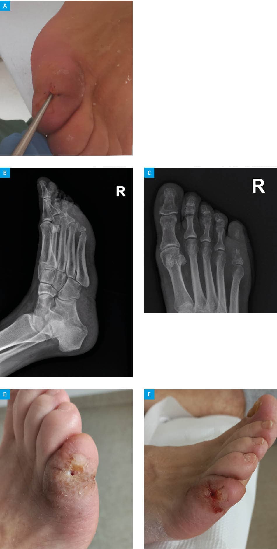
Intensive insulin therapy was started, along with continuous glucose monitoring, diabetes education, and wound care (surgical debridement, offloading, and systemic / local antibiotic therapy). Acinetobacter pittii was cultured from the wound (Figure 1D and 1E).2 The wound healed within 3 weeks, and amputation was avoided. Due to persistent dyslipidemia, statin therapy was initiated. Following optical coherence tomography, the patient was qualified for intravitreal anti–vascular endothelial growth factor therapy for diabetic macular edema.
The patient’s diabetic foot syndrome was neuropathic and likely due to mechanical trauma. Contributing factors included chronic hyperglycemia, smoking, and dyslipidemia. Early treatment prevented amputation. The lack of an earlier diabetes diagnosis, despite the presence of risk factors, might have contributed to the rapid onset and progression of chronic complications.3
This case highlights the diagnostic challenges associated with diabetes classification. The patient’s normal body weight and moderately low C‑peptide levels were suggestive of T1D, while her middle age at onset, dyslipidemia, multiple complications at diagnosis, family history of T2D, absence of signs of absolute insulin deficiency, and suspected gestational diabetes pointed toward T2D.4 Despite the negative results of antibody tests (approximately in 5%–10% of newly diagnosed white Europeans), which do not rule out an autoimmune process, a diagnosis of T1D was ultimately made, which is highly important, as its treatment fundamentally relies on insulin therapy.5
- Holt RIG, DeVries JH, Hess‑Fischl A, et al. The management of type 1 diabetes in adults. A consensus report by the American Diabetes Association (ADA) and the European Association for the Study of Diabetes (EASD). Diabetologia. 2021; 64: 2609‑2652. | Crossref
- Lipsky BA, Senneville É, Abbas ZG, et al. Guidelines on the diagnosis and treatment of foot infection in persons with diabetes (IWGDF 2019 update). Diabetes Metab Res Rev. 2020; 36 (Suppl 1): e3280. | Crossref
- Araszkiewicz A, Borys S, Broncel M, et al. Standards of care in diabetes. The position of Diabetes Poland ‑ 2025. Curr Top Diabetes. 2025; 5: 1‑158. | Crossref
- Możdżeń K, Pośpiech JP, Murawska AJ, et al. „Double diabetes?” Diagnostic and therapeutic challenges in patient with established type 1 diabetes mellitus, developing features of type 2 diabetes mellitus. Pol Arch Intern Med. 2025; 135: 16991. | Crossref
- Tridgell DM, Spiekerman C, Wang RS, Greenbaum CJ. Interaction of onset and duration of diabetes on the percent of GAD and IA‑2 antibody‑positive subjects in the type 1 diabetes genetics consortium database. Diabetes Care. 2011; 34: 988‑993. | Crossref
ARTICLE INFORMATION