Toxic epidermal necrolysis as a rare complication of pembrolizumab immunotherapy in the treatment of squamous cell lung cancer

 CC BY 4.0
Toxic epidermal necrolysis as a rare complication of pembrolizumab immunotherapy in the treatment of squamous cell lung cancer
Toxic epidermal necrolysis (TEN; Lyell syndrome) and Stevens–Johnson syndrome (SJS) are a continuum of severe, life‑threatening drug reactions involving separation of the epidermis, erythematous or purpuric lesions, skin blisters, and mucosal erosions, resembling severe burns. The abovementioned syndromes are differentiated based on the proportion of the body surface area (BSA) affected by the lesions, that is, where SJS affects less than 10% of the BSA and TEN over 30%, the involvement of 10%–30% of the BSA is defined as SJS/TEN overlap.1
A 65‑year‑old man with generalized squamous cell carcinoma of the left lung was admitted to a pulmonology, allergology, and internal medicine department due to pneumonia and swallowing disorders. Physical examination showed extensive erythematous and edematous lesions, mainly on the abdomen, back, and buttocks, along with erosions on the oral mucosa. Within 24 hours, the lesions began to coalesce and spread to the extremities and other mucosal sites. Subsequently, blisters developed (Figure 1A and 1B), followed by erosions and progressive epidermal detachment (Figure 1C–1E). The feet were the last to develop epidermal sloughing (Figure 1F). Ultimately, according to the Wallace rule of nines2, the eruptions were estimated to cover approximately 35% of the BSA, including the abdomen, back, buttocks, perineum, feet, and hands, with isolated lesions on the upper and lower extremities. Painful erosive and desquamative lesions involved the oral and urethral mucosa (Figure 1G), while the conjunctiva showed an ocular surface epithelial defect. The patient had persistent fever throughout hospitalization. Yet, blood and urine cultures were negative, and his general condition remained relatively good. The patient was eventually diagnosed with TEN triggered by pembrolizumab. The first symptoms appeared approximately 5 days after the first cycle of pembrolizumab treatment (erythematous and edematous lesions of the trunk, pain, and swelling in the mouth and throat). Significant local deteriorations of the skin condition occurred approximately 10 days after drug administration. This timeline is consistent with the literature reporting that SJS/TEN most frequently occurs during the first treatment cycle, typically between 2 and 180 days after drug administration (median, 15 d).3
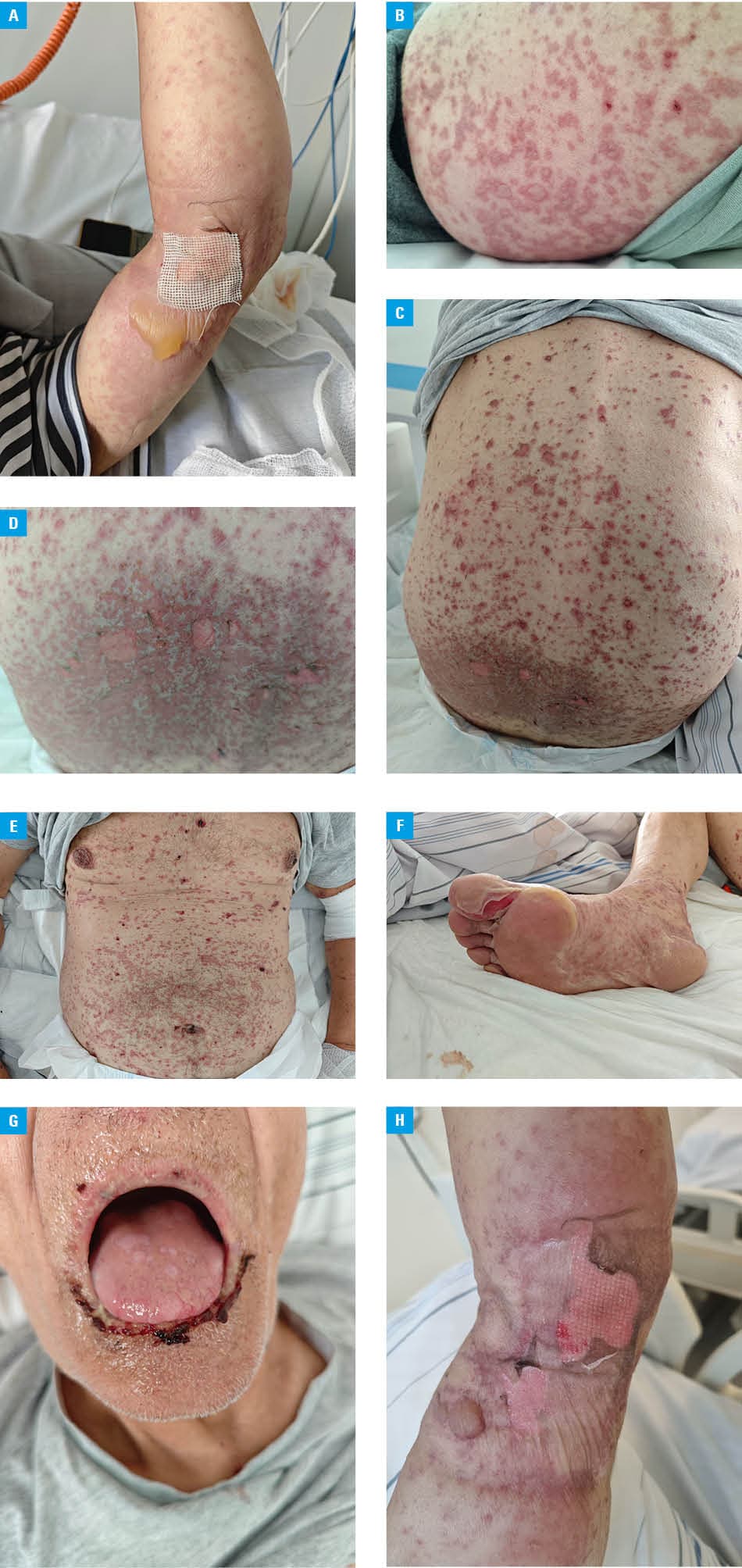
The treatment involved intravenous administration of methylprednisolone (daily dose of 1 mg/kg body weight [bw]) and intravenous immunoglobulins (total dose of 3 g/kg bw), given over 5 days. Clobetasol propionate ointment was applied topically to the erythematous lesions. For the ocular lesions, moisturizing preparations, dexamethasone, cyclosporine, and tobramycin were applied in the form of eye drops. Treatment of the lesions in the oral cavity included topical application of preparations containing nystatin, lidocaine, and hydrocortisone in the form of rinses. Wound care involved disinfection and application of paraffin dressings. Large blisters were punctured, leaving the epidermis as a natural biological dressing (Figure 1A and 1H). Supportive care included fluid therapy, pain control, and gradual transition from parenteral to oral nutrition. The patient remained under the care of a multidisciplinary team consisting of allergists, dermatologists, internists, otolaryngologists, and ophthalmologists. Due to pneumonia, antibiotic therapy was implemented in accordance with the local data on microbial resistance. The patient was discharged in a good general condition, with regressing lesions and several days of ongoing re‑epithelialization.
Pembrolizumab, a humanized anti‑programmed death (PD) 1 monoclonal antibody which blocks the interaction between the PD‑1 receptor and PD ligands 1 and 2, has been identified as a potential cause of TEN. It enhances the response of T lymphocytes and activates immune mechanisms, including anti‑tumor mechanisms.4 The drug is primarily used to treat melanoma, lung, head, neck, stomach, and cervical cancers, as well as Hodgkin lymphoma. According to the German Association of the Scientific Medical Societies guidelines,5 pembrolizumab is classified as “under observation” in terms of its potential to cause TEN, which means that isolated cases have been reported in the literature, and epidemiological data remain inconclusive. Available analyses confirm that SJS and TEN are rare but potentially fatal complications of treatment with pembrolizumab. A systematic review by Zhou et al6 found only a few case reports in the literature.
- Bastuji‑Garin S, Rzany B, Stern RS, et al. Clinical classification of cases of toxic epidermal necrolysis, Stevens–Johnson syndrome, and erythema multiforme. Arch Dermatol. 1993; 129: 92‑96. | Crossref
- Wallace AB. The exposure treatment of burns. Lancet. 1951; 1: 501‑504. | Crossref
- Wu Z, Li X, Huang R, et al. Clinical features, treatment, and prognosis of pembrolizumab‑induced Stevens‑Johnson syndrome / toxic epidermal necrolysis. Invest New Drugs. 2025; 43: 74‑80. | Crossref
- Qu J, Wang L, Jiang M, et al. A review about pembrolizumab in first‑line treatment of advanced NSCLC: focus on KEYNOTE studies. Cancer Manag Res. 2020; 12: 6493‑6509. | Crossref
- Heuer R, Paulmann M, Nast A, et al. Diagnosis and therapy of epidermal necrolysis (Stevens‑Johnson syndrome and toxic epidermal necrolysis) ‑ Langversion 1.1. AWMF‑Register No. 013‑103. 2024 [in German]. https://register.awmf.org/assets/guidelines/013‑103l_S3_Diagnostik‑Therapie‑epidermale‑Nekrolyse‑Stevens‑Johnson‑Syndrom‑toxisch‑epidermale‑Nekrolyse‑SJS‑TEN_2024‑11.pdf. Accessed August 21, 2025
ARTICLE INFORMATION