A 65‑year‑old man was admitted to a cardiac surgery department with massive pulmonary embolism and severe right ventricular failure. The patient did not present any symptoms that would suggest ischemia of the central nervous system or peripheral embolism, except for those related to pulmonary circulation. Transesophageal echocardiography showed a mobile, ballooning structure with the morphology of a thrombus in the right atrium, passing through the patent foramen ovale into the left atrium (Figure 1A and 1B). The structure in both the right and left atrium was approximately 40 mm in length. Thanks to the use of 3‑dimensional (3D) echocardiography, especially photorealistic imaging, it was possible to accurately assess the morphology of the structure (Figure 1C and 1D).1,2 The thrombus was removed from the fossa ovalis region (Figure 1E and 1F). By comparing the extracted thrombus with the preoperative 3D echocardiographic image, we ensured its complete removal. In addition, surgical thrombectomy of massive thrombi in the right and left pulmonary arteries was performed. After disconnecting extracorporeal circulation, a significant improvement in right ventricular contractility was observed, and no additional structures were identified in the heart chambers.
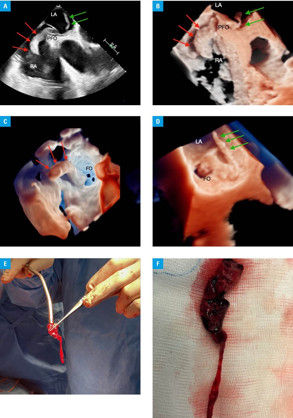
Abbreviations: FO, foramen ovale; LA, left atrium; PFO, patent foramen ovale; RA, right atrium
- Cheng K, Monaghan MJ, Kenny A, et al. 3D echocardiography: benefits and steps to wider implementation. Br J Cardiol. 2018; 25: 63‑68. | Crossref
- Lang RM, Badano LP, Tsang W, et al. EAE/ASE recommendations for image acquisition and display using three‑dimensional echocardiography. Eur Heart J Cardiovasc Imaging. 2012; 13: 1‑46. | Crossref
ARTICLE INFORMATION