
Fulminant periannular destruction after valve-in-valve transcatheter aortic valve implantation unmasking advanced HIV infection

 CC BY 4.0
Fulminant periannular destruction after valve-in-valve transcatheter aortic valve implantation unmasking advanced HIV infection
CC BY 4.0
Structural valve deterioration (SVD) is a recognized limitation of bioprostheses, often manifesting 10–15 years postimplantation.1 Valve‑in‑valve transcatheter aortic valve implantation (ViV‑TAVI) is increasingly utilized for failed bioprostheses in patients at an elevated risk of surgery.2 Infective endocarditis (IE) following TAVI, albeit uncommon, carries grave prognostic implications and complex diagnostic / therapeutic challenges.3
A 61‑year‑old man, 11 years after surgical AV replacement (Perimount Magna 25; Edwards Lifesciences LLC, Irvine, California, United States) for severe aortic regurgitation, presented with progressive exertional dyspnea (New York Heart Association class III). Transthoracic echocardiography demonstrated advanced SVD with mixed dysfunction: peak / mean transvalvular gradients of 98/51 mm Hg and maximum flow velocity (Vmax) of 5 m/s, accompanied by mild regurgitation. The heart team adjudicated transfemoral ViV‑TAVI as the preferred strategy, and a self‑expanding Evolut PRO 26 valve (Medtronic, Dublin, Ireland) was deployed without intraprocedural complications (Figure 1A).
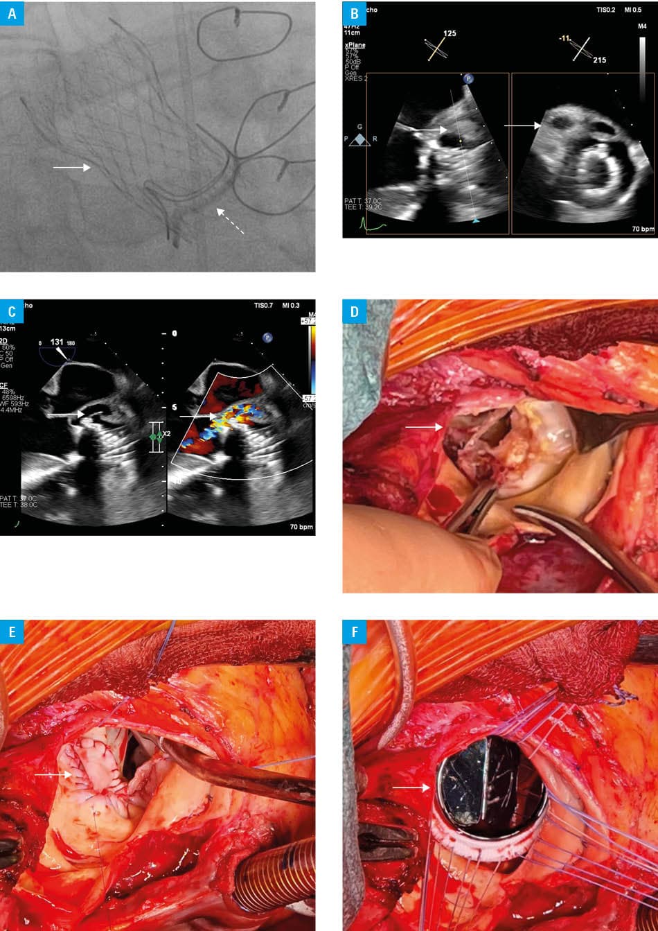
Within days, the patient developed fever with rising inflammatory indices. Transesophageal echocardiography (TEE) 1 week postoperatively identified an approximately 20‑mm periannular abscess at the aortomitral curtain with mild paravalvular regurgitation (PVR; Figure 1B). Repeat TEE 1 week later showed enlargement to a 25 mm × 25 mm cavity with a new aortocavitary fistula and moderate PVR (Figure 1C). After 2 weeks, TEE demonstrated a 34‑mm cavity with severe PVR (estimated regurgitant volume, 130 ml). All blood cultures remained sterile, and targeted serology for fastidious pathogens and fungal blood cultures were negative. A working diagnosis of prosthetic valve IE was established based on the 2023 Duke criteria (major imaging criterion with supportive minors).4 Intravenous antimicrobial therapy was initiated according to the local IE protocol.
Concomitantly, screening for immunodeficiency was undertaken. Fourth‑generation HIV screening yielded a positive result, which was confirmed by nucleic acid testing, demonstrating high plasma HIV RNA level. It should be emphasized that, prior to this, the patient had no clinical features of immunosuppression, no documented HIV testing, and no reported risk behaviors or other epidemiologic risk factors. Immunophenotyping demonstrated profound CD4+ T‑cell depletion (absolute CD4 count, 74/µl; reference range [RR], 300–1400/µl; 6% of lymphocytes) with marked CD8+ expansion (79%; 1025/µl; RR, 200–900/µl) and a severely inverted CD4/CD8 ratio (approximately 0.07; RR, 0.5–2.5), fulfilling immunological criteria for advanced HIV infection. Combination antiretroviral therapy, consisting of a dolutegravir‑based and tenofovir‑containing regimen, was initiated promptly within a multidisciplinary framework with infectious disease specialists.
Due to rapid clinical deterioration with hemodynamic compromise, the patient was referred for urgent redo surgery. Intraoperative findings confirmed extensive periannular destruction with an abscess cavity and loss of continuity localized in the noncoronary sinus (Figure 1D). TAVI frame and the degenerated surgical bioprosthesis were explanted. Radical debridement was performed, and the defect was reconstructed with a bovine pericardial patch (Figure 1E). Given extensive tissue loss and annular discontinuity, a mechanical valve (On‑X 25; On‑X Life Technologies, Austin, Texas, United States) was implanted to maximize durability and minimize the risk of another intervention (Figure 1F). The postoperative course was uneventful with satisfactory wound healing. Intravenous antimicrobial therapy was continued and subsequently transitioned to microbiology / infectious disease guidance; antiretroviral therapy was maintained.
Three months after discharge, the patient remained clinically stable. Transthoracic echocardiography showed preserved left ventricular ejection fraction (50%), optimal mechanical valve hemodynamics (peak / mean gradient, 10/6 mm Hg; Vmax, 1.6 m/s) and absence of paravalvular regurgitation.
This case illustrates post‑TAVI IE with fulminant periannular spread involving the aortomitral curtain, resulting in catastrophic valvular dysfunction. TEE is pivotal for early recognition of periannular extension and surgical planning. Post–ViV‑TAVI follow‑up mirrored contemporary TAVI protocols: TTE at discharge / in 30 days or less, at 6 and 12 months, and annually thereafter.5 In our patient, emerging clinical signs of infection prompted early TEE, enabling timely identification of the pathology. Given the evolving HIV epidemiology, with late diagnoses still common, cross‑specialty vigilance is essential. Timely, routine HIV testing in atypical or severe infections may expedite life‑saving therapy. A multidisciplinary team is critical for timely decision‑making in catastrophic prosthetic valve IE, coordinating surgery timing, antimicrobial therapy, and combination antiretroviral therapy.
- Bourguignon T, Bouquiaux‑Stablo A‑L, Candolfi P, et al. Very long‑term outcomes of the Carpentier–Edwards Perimount valve in aortic position. Ann Thorac Surg. 2015; 99: 831‑837. | Crossref
- Dvir D, Webb JG, Bleiziffer S, et al. Transcatheter aortic valve implantation in failed bioprosthetic surgical valves. JAMA. 2014; 312: 162‑170. | Crossref
- Del Val D, Panagides V, Mestres CA, et al. Infective endocarditis after transcatheter aortic valve replacement: JACC state‑of‑the‑art review. J Am Coll Cardiol. 2023; 81: 394‑412. | Crossref
- Fowler VG Jr, Durack DT, Selton‑Suty C, et al. The 2023 Duke‑International Society for Cardiovascular Infectious Diseases criteria for infective endocarditis: updating the modified Duke criteria. Clin Infect Dis. 2023; 77: 518‑526. | Crossref
- Vahanian A, Beyersdorf F, Praz F, et al. 2021 ESC/EACTS guidelines for the management of valvular heart disease. Eur Heart J. 2022; 43: 561‑632. | Crossref
ARTICLE INFORMATION