
From leg swelling to life-threatening emergency: spontaneous iliac vein rupture associated with May–Thurner syndrome

 CC BY 4.0
From leg swelling to life-threatening emergency: spontaneous iliac vein rupture associated with May–Thurner syndrome
CC BY 4.0
Spontaneous iliac vein rupture (SIVR) is a rare but life‑threatening complication of acute iliofemoral deep vein thrombosis (DVT). Its detection and treatment are challenging and, with only case reports available, raising physician awareness is crucial.1
A 63‑year‑old woman without prior trauma presented with rapidly progressing edema, erythema, and severe pain of the left lower limb. Right before admission, she experienced abdominal pain and a brief loss of consciousness. Anamnesis for cardiorespiratory disease, cancer, and thrombophilia was negative. Physical examination showed absent distal pulses, impaired sensation and mobility, and cutaneous tension—findings consistent with phlegmasia cerulea dolens. Laboratory workup showed markedly elevated levels of D‑dimer (>4000 ng/ml; reference range [RR] <500 ng/ml) and C‑reactive protein (105.8 mg/l; RR <10 mg/l), leukocytosis (leucocytes, 22.61 × 10³/μl; RR, 4–10 × 103/μl), hyperlactatemia (lactate, 8.9 mmol/l; RR, 0.5–1.6 mmol/l), extremely elevated levels of myoglobin (67 953 ng/l; RR, 10–92 ng/ml) and creatinine (3 mg/dl; RR, 0.5–1 mg/dl), pH of 7.2, and severe hyperglycemia (glucose, 834 mg/dl; RR, 70–99 mg/dl). Ultrasound and computed tomography angiography (CTA) demonstrated extensive left‑side DVT from the left common iliac vein (LCIV) to the popliteal vein and a 4.5‑cm retroperitoneal hematoma (RH; Figure 1A and 1B). No arterial bleeding was identified, raising suspicion of SIVR. Uterine fibroids found on imaging were ruled out as unrelated. Following a multidisciplinary consultation, we diagnosed massive iliofemoral DVT complicated by RH, new‑onset diabetic ketoacidosis (DKA), acute kidney injury (AKI), and rhabdomyolysis (likely involving the muscles of the leg and the iliopsoas muscle).
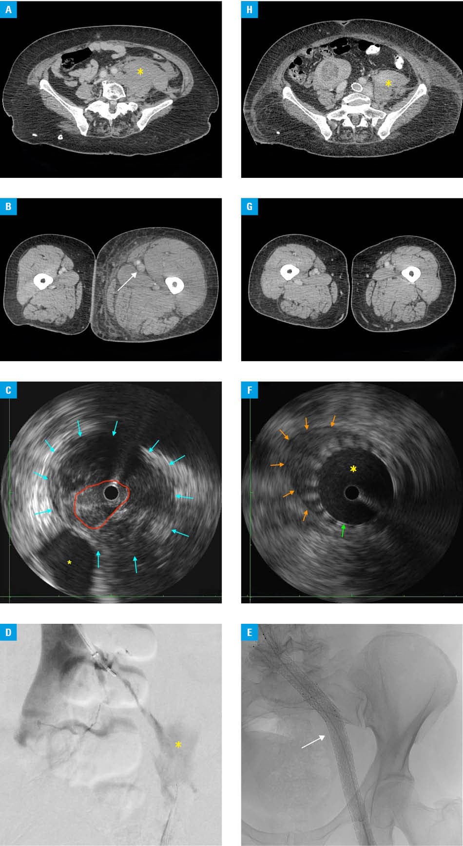
Initial stabilization included administration of unfractionated heparin, aggressive fluid therapy with electrolyte correction, bed rest with limb elevation, and a continuous insulin infusion. Despite treatment, AKI progressed to anuria, requiring urine alkalinization with bicarbonate. After 3 days of heparin, the hemoglobin level decreased from 12.1 to 9.6 g/dl (RR, 12–16 g/dl).
Based on the clinical presentation and imaging findings suggestive of SIVR, at a joint clinical‑radiological meeting, endovascular intervention was selected as preferable due to the available methods and the unit’s experience.
Vascular access was obtained via the right common femoral vein (FV), the occluded left common FV (LCFV), and the right internal jugular vein. An inferior vena cava OptionElite filter (Argon Medical Devices Inc., Frisco, Texas, United States) was placed. Following recanalization of the left iliac axis, venography and intravascular ultrasound confirmed SIVR and May–Thurner syndrome (MTS; Figure 1C and 1D). Aspiration thrombectomy was carried out using a Lightning 12F catheter (Penumbra Inc., Alameda, California, United States). Angioplasty of the left deep FV, LCFV, left external IV, and LCIV was performed with 12‑mm and 14‑mm balloons (Becton, Dickinson and Company, Tempe, Arizona, United States). Venovo stents (14 mm × 160 mm and 14 mm × 100 mm; Becton, Dickinson and Company) were implanted with mandatory postdilatation. Stent positioning and patency were confirmed (Figure 1E). Postprocedurally, therapeutic enoxaparin at a dose of 80 mg twice daily was initiated. AKI and rhabdomyolysis improved markedly with treatment, and limb symptoms resolved. At discharge, compression stockings and 20 mg of rivaroxaban daily were prescribed. The inferior vena cava filter was retrieved 4 weeks later. Follow‑up CTA and intravascular ultrasound showed RH regression and patent stents (Figure 1F–1H). The patient was uneventful at 10‑month follow‑up.
SIVR is an exceptionally rare but life‑threatening cause of RH, with fewer than 100 cases reported. The condition predominantly affects middle‑aged and elderly women with a concomitant left‑sided DVT. Mortality rates reach up to 22%, making early recognition crucial.1 SIVR frequently occurs in association with MTS, where chronic compression of the IV weakens the venous wall and predisposes to rupture during acute DVT.2
However, the overall etiology of SIVR remains poorly understood, with previous literature speculating about additional risk factors, such as a loss of estrogen effect or sudden increase in venous pressure due to Valsalva maneuvers (eg, heavy lifting or defecation).1,2
Severe AKI likely resulted from synergistic factors: rhabdomyolysis‑induced nephrotoxicity, systemic hypoperfusion from retroperitoneal bleeding, and DKA‑related dehydration exacerbating renal ischemia.3 MTS, combined with hypercoagulability in DKA and systemic inflammation, explains the rapid progression to phlegmasia cerulea dolens.4
Management must balance hemorrhage control with anticoagulation to prevent further thrombosis and possible pulmonary embolism. Open surgery was historically the most common approach due to delayed diagnosis. However, endovascular treatment (EVT) offers better outcomes, may prevent post‑thrombotic syndrome, and achieve long‑term venous patency in MTS. Conservative treatment can be considered when RH is contained and the patient remains stable with no severe phlegmasia symptoms.1
In SIVR, RH with a mass effect compresses the vein, raising pressure at the rupture site and worsening bleeding, while intimal injury may further narrow the lumen. Unlike high‑pressure arterial bleeding, where sealing the defect is the priority, in venous rupture, the main goal is to restore venous outflow and lower the pressure at the rupture site.5 We opted for widely available bare stents, because they maintain collateral perfusion and have a lower risk of in‑stent thrombosis. Covered stents may occlude the internal IV and other confluences, compromising venous return and future intervention options.
This case illustrates the diagnostic and therapeutic challenges of SIVR and a vicious cycle of metabolic complications, safely managed conservatively and with EVT. Early diagnosis enables EVT with excellent survival, while delayed diagnosis often necessitates emergency surgery that carries a risk of substantial mortality. Multidisciplinary coordination is essential.
- Kooiman AL, Bakas JM, Van Fessem JMK, et al. Spontaneous iliac vein ruptures: a systematic review. Vasc Endovascular Surg. 2023; 57: 617‑625. | Crossref
- Nishimoto Y, Toma M, Iwasa K, Sato Y. Successful staged management of a spontaneous iliac vein rupture associated with May–Thurner syndrome: a case report. Eur Heart J Case Rep. 2021; 5: ytab316. | Crossref
- Hebert JF, Burfeind KG, Malinoski D, Hutchens MP. Molecular mechanisms of rhabdomyolysis‑induced kidney injury: from bench to bedside. Kidney Int Rep. 2023; 8: 17‑29. | Crossref
- Domingueti CP, Dusse LMS, Carvalho MDG, et al. Diabetes mellitus: the linkage between oxidative stress, inflammation, hypercoagulability and vascular complications. J Diabetes Complications. 2016; 30: 738‑745. | Crossref
- Cha JG, Lee SY, Hong J, et al. Traumatic iliac vein rupture managed using a bare‑metal stent. Trauma Case Rep. 2022; 37: 100589. | Crossref
ARTICLE INFORMATION