
Integrated imaging and cellular bioenergetic profiling of the aorta in a patient with Loeys–Dietz syndrome

 CC BY 4.0
Integrated imaging and cellular bioenergetic profiling of the aorta in a patient with Loeys–Dietz syndrome
CC BY 4.0
Loeys–Dietz syndrome (LDS) is a rare, autosomal dominant connective tissue disorder caused by pathogenic variants in the genes encoding proteins in the transforming growth factor β signaling pathway. This disorder predisposes to severe vascular complications, including aneurysms and aortic dissections, often requiring early surgical management.1 Beyond structural vascular lesions, LDS also involves mitochondrial and cytoskeletal dysfunction in vascular wall cells. These abnormalities may contribute to impaired endothelial homeostasis and pathological remodeling of the aortic wall.2,3
A 34‑year‑old man with LDS type 3 with heterozygous potentially pathogenic (ACMG class 4) variant NM_005902.4(SMAD3):c.527_528del, p.Ile176ThrfsTer11 (chr15‑67457716ATT>A) was referred to our university hospital for surgical treatment due to significant dilatation of the aortic root (55 mm on transthoracic echocardiography). On computed tomography angiography of the head, chest, abdomen, and pelvis, no other vascular lesions were found, apart from the aortic root dilatation (Figure 1A). The patient had a history of asthma and scoliosis but no previous cardiovascular interventions.
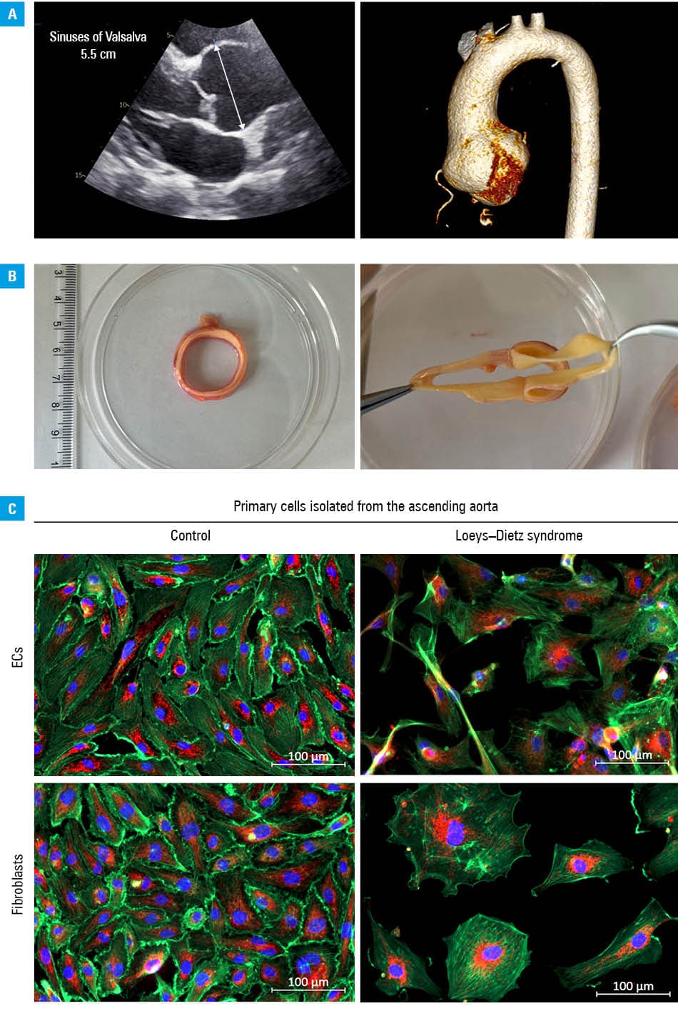
The patient underwent the David procedure, a valve‑sparing aortic root repair. The excised aortic wall segment (Figure 1B) was immediately examined to explore the cellular phenotype associated with LDS. We performed ex vivo analysis of LDS patient‑derived endothelial cells (ECs) and fibroblasts, and compared them with control cells isolated from the ascending aorta of a 67‑year‑old woman with coronary artery disease, removed during coronary artery bypass graft surgery. After enzymatic dissociation of the internal and adventitial aortic layers and subsequent magnetic separation, both cell types were cultured under standard conditions. The intimal ECs derived from control and LDS patients’ aorta gradually formed a confluent monolayer, exhibiting the typical cobblestone appearance. Control adventitial fibroblasts displayed an elongated spindle‑shaped morphology, and those derived from the LDS patient showed a more heterogenous range of shapes. The cultured cells were examined using light and fluorescence microscopy (Axio Observer 7 inverted microscope; Carl Zeiss, Oberkochen, Germany).4 Mitochondrial distribution (translocase of the outer mitochondrial membrane 20 staining) and cytoskeletal organization (phalloidin staining) were visualized to assess cellular bioenergetics and structural integrity. This combined morphological and subcellular imaging enabled a direct assessment of vascular cell condition in relation to the patient’s clinical phenotype.
Under evaluation, clear differences in cell structure were observed between the LDS‑derived cells and control ones (Figure 1C; Supplementary material, Figures S1 and S2). Control ECs formed a uniform, organized monolayer with evenly distributed mitochondria and dense actin filaments. In the patient with LDS, ECs exhibited a preserved cytoskeletal framework, although subtle mitochondrial redistribution was noted. In contrast, LDS‑derived fibroblasts demonstrated marked abnormalities, including elongated morphology, perinuclear mitochondrial clustering, disturbed actin organization, and absence of adhesive connections with the neighboring cells.
This case illustrates that integrating clinical imaging with ex vivo cellular profiling can show novel aspects in vascular pathologies. By linking clinical presentation, imaging findings, and cellular morphology and integrity, such a multimodal approach provides a translational perspective on the disease process that extends beyond conventional anatomical imaging.
From a clinical standpoint, this approach may support the development of targeted therapies aimed at restoring endothelial energy balance and preventing aneurysm progression in disorders such as LDS. Pharmacological modulation of mitochondrial function using, for instance, sodium‑glucose cotransporter 2 inhibitors, has shown potential benefits in improving endothelial bioenergetics and nitric oxide availability.5
In summary, this report demonstrates that enriching cardiovascular imaging with patient‑derived cellular analyses improves our understanding of the mechanisms underlying vascular fragility in hereditary genetic aortic diseases, such as LDS, and supports the search for personalized therapeutic strategies.
- MacCarrick G, Black JH 3rd, Bowdin S, et al. Loeys–Dietz syndrome: a primer for diagnosis and management. Genet Med. 2014; 16: 576‑587. | Crossref
- Verstraeten A, Fedoryshchenko I, Loyes B. The emerging role of endothelial cells in the pathogenesis of thoracic aortic aneurysm and dissection. Eur Heart J. 2023; 44: 1262‑1264. | Crossref
- Marcos‑Rios D, Rochano‑Ortiz A, San Sebastián‑Jaraba I. Mitochondrial dysfunction: a new hallmark in hereditable thoracic aortic aneurysm development. Cells. 2025; 14: 618. | Crossref
- Braczko A, Harasim G, Kawecka A, et al. Blocking cholesterol formation and turnover improves cellular and mitochondria function in murine heart microvascular endothelial cells and cardiomyocytes. Front Physiol. 2023; 14: 1216267. | Crossref
- Walczak I, Braczko A, Paterek A, et al. Dapagliflozin, an SGLT2 inhibitor, improves endothelial cell energy metabolism through enhanced mitochondrial respiration. Cell Physiol Biochem. 2025; 59: 235‑251. | Crossref
SUPPLEMENTARY MATERIAL
ARTICLE INFORMATION