

 CC BY 4.0
A 36‑year‑old woman was admitted to an emergency department (ED) following out‑of‑hospital cardiac arrest due to malignant ventricular arrhythmias. She was successfully resuscitated by paramedics. On arrival, she was conscious, hemodynamically stable (blood pressure, 111/80 mm Hg; oxygen saturation, 100%; heart rate, 93 bpm). Electrocardiogram (ECG) showed sinus rhythm at 100 bpm, PQ interval of 160 ms, QRS duration of 90 ms, QT interval of 366 ms, and corrected QT interval (QTc) of 422 ms.
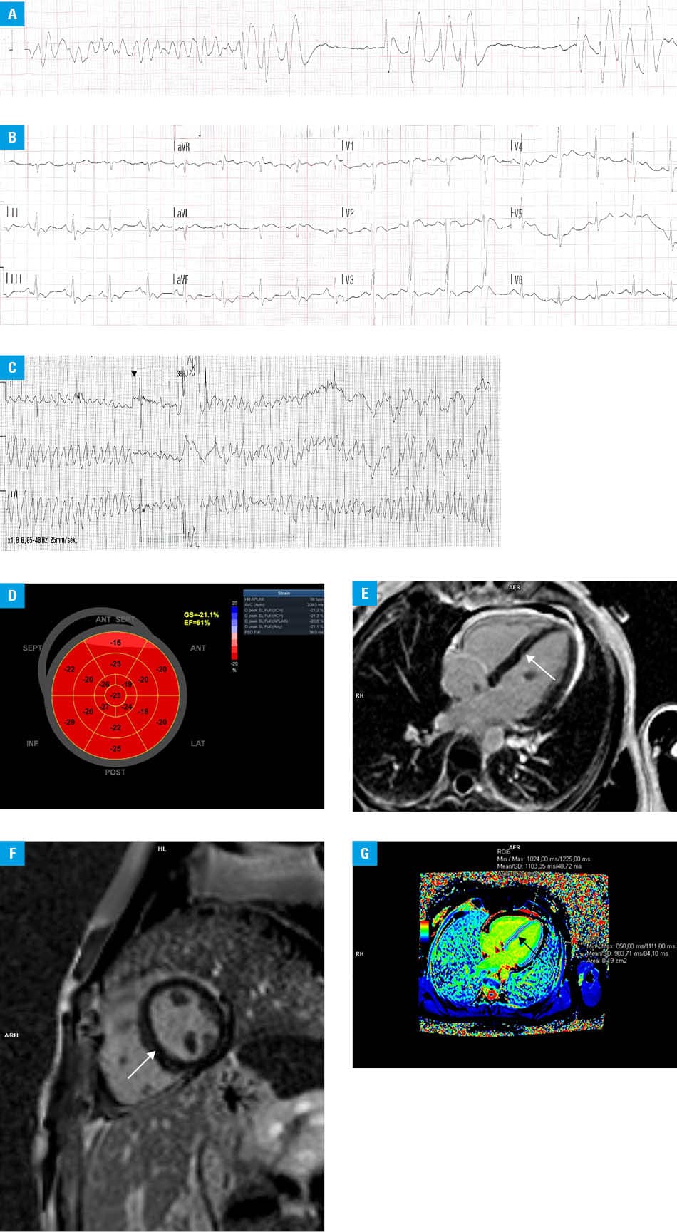
A syncopal episode, preceded by severe burning chest pain and emesis, occurred 30 minutes after sumatriptan ingestion. The patient briefly regained consciousness before collapsing again in front of paramedics. Ventricular fibrillation (VF) was documented (Figure 1A), and return of spontaneous circulation (ROSC) was achieved after 3 defibrillation shocks and cardiopulmonary resuscitation (CPR). Postresuscitation ECG showed sinus rhythm with diffuse ST‑segment depressions and a QTc interval of 382 ms (Figure 1B). During transportation, the patient experienced polymorphic ventricular tachycardia (torsade de pointes [TdP]), leading to another cardiac arrest (Figure 1C). Three additional defibrillation attempts and CPR resulted in ROSC.
The patient had a 19‑year history of using sumatriptan for migraines, with no other diagnosed medical conditions. Similar but milder episodes, lasting approximately 20 minutes and alleviated by vomiting, had occurred recently. She smoked 40 cigarettes per day (40 pack‑years) and consumed approximately 2 liters of energy drinks daily (approximately 500 mg of caffeine). She denied recent alcohol or psychoactive substance use, as well as allergies or pregnancy. Her mother had died suddenly at the age of 30 years.
Initial evaluation performed on admission to the ED included ECG, laboratory workup, echocardiography, and computed tomography for pulmonary embolism. Laboratory findings were as follows: potassium level of 3.3 mmol/l (reference range [RR], 3.5–5 mmol/l), C‑reactive protein level within the normal range (<5 mg/l); arterial pH of 7.24 (RR, 7.35–7.45), and lactate level of 7 mmol/l (RR, 0.5–1.6 mmol/l). The level of high‑sensitivity troponin T was initially within the normal range (13.4 ng/l; RR <14 ng/l), later increasing to 85.8 ng/l. Initial echocardiography showed normal chamber sizes, mildly reduced left ventricular ejection fraction (LVEF; 45%), global hypokinesis, and no valvular disease or pericardial effusion. Coronary angiography showed normal coronary arteries.
LVEF later improved to 61% (Figure 1D). Hypokinesis of the apical segment of the interventricular septum (IVS) persisted. Treatment included potassium, magnesium, and metoprolol at a dose of 50 mg/day. No further arrhythmia episodes occurred during hospitalization.
Cardiac magnetic resonance imaging (MRI) visualized linear mid‑wall late gadolinium enhancement in the mid and apical IVS (Figure 1E and 1F) and prolonged T1 relaxation time (Figure 1G), consistent with nonischemic origin of myocardial fibrosis.1 Genetic tests involving part of the currently known cardiomyopathy‑related mutations in specific genes yielded negative results for pathogenic mutations. Migraine treatment was modified to rimegepant at a dose of 75 mg ad hoc.
A single‑chamber implantable cardioverter‑defibrillator (ICD) was implanted. Electrophysiological study during the procedure showed low signal amplitudes (midseptum sensing, 3 mV) in the IVS, consistent with MRI findings. At 6‑month follow‑up, the patient was asymptomatic, arrhythmia‑free, and had quit smoking and energy drinks.
This case highlights the complexity of diagnostic evaluation in a young patient with significant cardiac risk factors, who presented with chest pain following sumatriptan intake, ultimately leading to cardiac arrest. Sumatriptan may be cardiotoxic by prolonging the QTc interval2 or via coronary vasospasm mediated by the activation of 5‑hydroxytryptamine 1B/1D receptors (present in both cerebral and coronary arteries).3
Ventricular arrhythmias following sumatriptan use are rare and documented mainly in isolated case reports. Real‑world pharmacovigilance databases provide signals of risk but lack precise incidence data. More evidence is available regarding other symptoms, such as chest pain and tightness, and an increased risk of coronary vasospasm (approximately 5‑fold, as compared with nonusers).4 Women report sumatriptan‑related adverse effects about 3 times more frequently than men.4
While sumatriptan may have acted as the immediate trigger for VF–TdP in our case, the diagnostic pathway required consideration of differential diagnoses and potential confounders. The hypokalemia observed on admission may have had a bidirectional relationship with the cardiac arrest: it could have contributed to the development of ventricular arrhythmia, but it could also have been a consequence of TdP with subsequent cardiac arrest and adrenergic stimulation, all of which can promote an intracellular shift of potassium and thereby lead to electrolyte imbalance. Invasive testing for vasospastic angina was not performed. Based on the cardiac MRI findings and family history, a decision was made to implant an ICD for secondary prevention of ventricular arrhythmias.
Given its potential cardiovascular adverse effects, initiation of sumatriptan therapy should be preceded by an assessment of cardiovascular risk. If any contraindications are identified, triptan therapy should be used with caution, or avoided altogether. Patient education regarding potential adverse effects, along with careful medication reconciliation, is also recommended to reduce the risk of potentially fatal arrhythmias.
- Petersen SE, Timperley J, Neubauer S. Left ventricular thrombi in a patient with left ventricular non‑compaction in visualisation of the rationale for anticoagulation. Heart. 2005; 91: e4. | Crossref
- Darpö B. Spectrum of drugs prolonging QT interval and the incidence of torsades de pointes. Eur Heart J Supp. 2001; 3: K70‑K80. | Crossref
- Longmore J, Maguire JJ, Macleod A, et al. Comparison of the vasoconstrictor effects of the selective 5‑HT 1D‑receptor agonist L‑775,606 with the mixed 5‑HT 1B/1D‑receptor agonist sumatriptan and 5‑HT in human isolated coronary artery. Br J Clin Pharmacol. 2000; 49: 126‑131. | Crossref
- Liu WH, Hu HM, Li C, et al. Real‑world study of adverse events associated with triptan use in migraine treatment based on the U.S. Food and Drug Administration (FDA) Adverse Event Reporting System (FAERS) database. J Headache Pain. 2024; 25: 206. | Crossref
ARTICLE INFORMATION