


 CC BY-NC-SA 4.0
Aortic stenosis is the most prevalent valvular heart disease. The number of transcatheter aortic valve implantation (TAVI) procedures is increasing as the population ages.1
A 90‑year‑old woman was admitted to a hospital after experiencing an episodic loss of consciousness. A diagnosis of acute coronary syndrome was made based on the patient’s symptoms and dynamic ST‑T wave changes on electrocardiography. Coronary angiography was urgently performed, but the results were negative. Transthoracic echocardiography (TTE) identified severe aortic stenosis, with a maximal pressure gradient of 158 mm Hg, mean pressure gradient of 91 mm Hg, and calculated aortic valve area of 0.3 cm2. The decision was made to perform urgent balloon aortic valve repair to stabilize the patient.2 The valve annulus parameters evaluated using computed tomography included a mean diameter of 25.7 mm (a minimum of 23 mm and a maximum of 28.4 mm). The area was measured at 521 mm2, with an area‑derived diameter of 25.8 mm. The perimeter was determined at 82.3 mm. The diameter derived from the perimeter was determined at 26.2 mm. This indicated that the optimal valve for the patient would be either CoreValve Evolut Pro 29 mm (Medtronic, Minneapolis, Minnesota, United States) or SAPIEN 26 mm (Edwards Lifesciences, Irvine, California, United States). Following 5 days of uneventful observation, TAVI was performed using a self‑expanding valve prosthesis CoreValve Evolut Pro 29 mm via left femoral access. Simultaneously, “chimney” stenting of the left main coronary artery was performed to mitigate the risk of coronary artery occlusion, given the markedly short distance (4 mm) between the left main coronary artery ostium and the aortic valve annulus (Figure 1A).
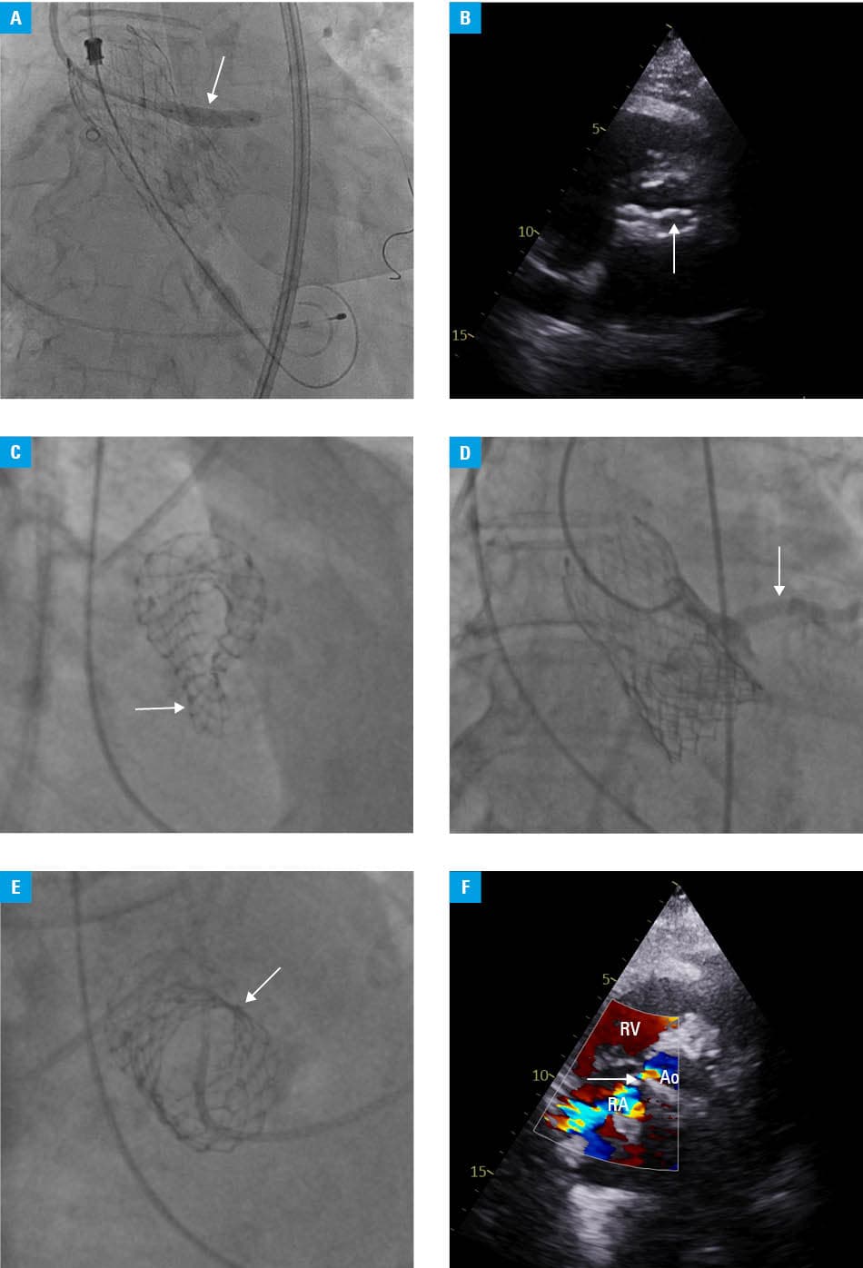
Abbreviations: Ao, aorta; RA, right atrium; RV, right ventricle
Within 24 hours postprocedure, the patient acutely deteriorated, presenting with sudden onset pulmonary edema. TTE showed an atypical, whetstone‑shaped bioprosthesis with signs of severe stenosis and significant regurgitation (Figure 1B). The patient was urgently transferred to a catheterization laboratory, where valve recoil was identified (Figure 1C). A series of balloon inflations (25 mm; Valver, Balton, Poland) was performed on the collapsed prosthesis (under TTE guidance to verify the intravalvular position), but it was not possible to restore an appropriate frame shape (Figure 1D). A bailout valve‑in‑valve procedure was performed, using a balloon expandable (BE) SAPIEN 3 Ultra valve prosthesis of 16 mm (Edwards Lifesciences),3 with a nominal 23‑ml volume postdilatation with a delivery system balloon (Commander; Edwards Lifesciences) due to regurgitation. This intervention yielded a favorable acute hemodynamic effect, as shown in Figure 1E, but TTE demonstrated presence of a large jet located between the aortic valve annulus and the right atrium (Figure 1F). The prevailing hypothesis for the etiology of this fistula was that it was most likely caused by an uncontrolled rupture of calcified tissues during postdilatation. Another potential mechanism for the development of the fistula, as reported in the existing literature, involves iatrogenic injury during TAVI, caused by the metallic frame of the bioprosthesis.4
In the following days, the patient showed no signs of heart failure and was deemed eligible for continued conservative management of the aorto‑atrial fistula.
The patient’s echocardiographic parameters were stable but other complications, including pneumonia, gastrointestinal bleeding, acute kidney injury requiring continuous hemodiafiltration, and critical limb ischemia, prolonged her hospital stay. The collection of multiorgan complications ultimately resulted in the patient’s death 14 days after the last interventional procedure.
There are 2 main types of valve prostheses dedicated for the TAVI procedure: self‑expanding (SE) and BE.5 The SE valve prosthesis exerts low radial force, requiring predilation in the calcified lesions. Rapid pacing is generally unnecessary, benefiting patients with low ejection fraction or hemodynamic instability. SE valves may be repositioned and carry a minimal risk of aortic annular injury. BE valves offer higher radial force, a reduced necessity for pre- and postdilation, and superior access to the coronary arteries in subsequent procedures. The risk of perforation is higher with BE valves, even when accounting for appropriate sizing of the prosthesis.
In the presented case, the adopted approach aligned with the prevailing consensus on the management of high‑risk patients. Regardless, the patient exhibited signs of prosthesis recoil following effective predilation. In addition, a fistula formed between the right heart chambers, despite adequate sizing of the BE prosthesis for the valve‑in‑valve procedure. Coronary calcium can be modified using atherectomy, lithotripsy, cutting, or ultra‑high‑pressure balloons. Conversely, TAVI relies exclusively on pre- and postdilation (a form of lithotripsy for the aortic valve is in the research phase). This often leaves the operator with a challenging decision when managing heavy calcifications. Management of the shunts depends on the symptomatology. If aneurysms are small, a conservative treatment approach, involving systematic echocardiographic follow‑up, can be effective. In the cases of larger aneurysms with elevated right ventricular systolic pressure and signs of right heart failure caused by overload, invasive repair—either percutaneous (with a vascular plug) or surgical—is indicated. The patient exhibited hemodynamic stability, and her subsequent complications were attributed to severe comorbidities. Consequently, a conservative treatment approach was implemented at that time.
- Pardo Sanz A, Zamorano Gómez JL. How to improve patient outcomes following TAVI in 2024? Recent advances. Pol Heart J. 2024; 82: 696‑701. | Crossref
- Tyczyński P, Chmielak Z, Dąbrowski M, et al. Elective versus rescue balloon aortic valvuloplasty for critical aortic stenosis. Kardiol Pol. 2020; 78: 982‑989. | Crossref
- Stąpór M, Trębacz J, Sobczyński R, et al. Transcatheter valve‑in‑valve aortic valve implantation for failed aortic homografts: a single‑center experience. Pol Heart J. 2025; 83: 492‑495. | Crossref
- Al Ayouby A, Gilard M, Hebert T, Didier R. Case report: iatrogenic left ventricular outflow tract to right atrium fistula after trans‑femoral transcatheter aortic valve implantation associated with asymmetric septal hypertrophy. Eur Heart J Case Rep. 2021; 5: ytab020. | Crossref
- Costa G, Criscione E, Reddavid C, Barbanti M. Balloon‑expandable versus self‑expanding transcatheter aortic valve replacement: a comparison and evaluation of current findings. Expert Rev Cardiovasc Ther. 2020; 18: 697‑708. | Crossref
ARTICLE INFORMATION