
Extensive deep vein thrombosis in the course of azygos continuation of the inferior vena cava treated with ultraslow low-dose thrombolysis

 CC BY 4.0
Extensive deep vein thrombosis in the course of azygos continuation of the inferior vena cava treated with ultraslow low-dose thrombolysis
CC BY 4.0
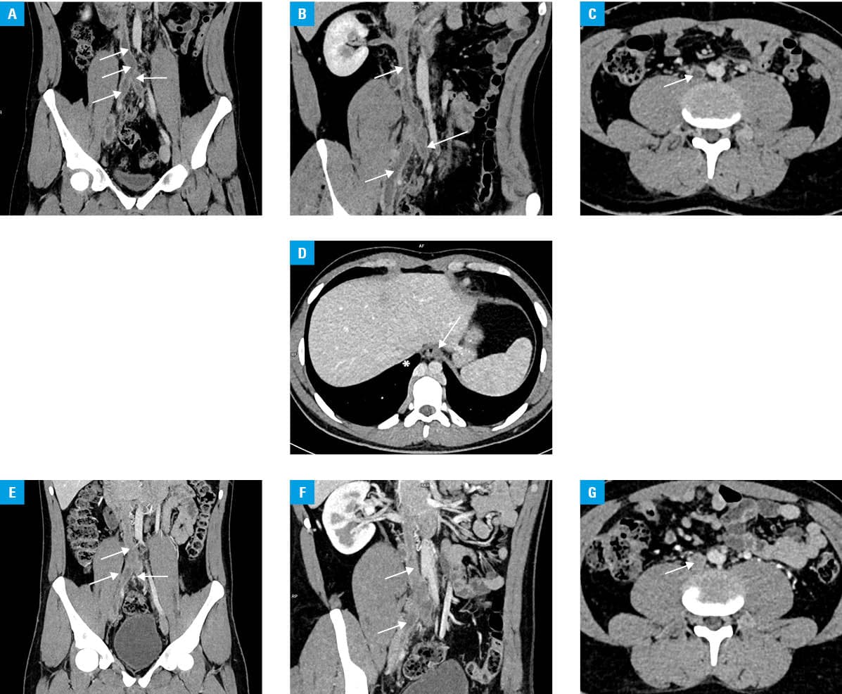
A 20‑year‑old man without prior medical history was admitted to an emergency department due to painful edema of the right lower extremity that had persisted for several days. Laboratory test results yielded a moderately increased C‑reactive protein concentration (11 mg/l; reference range [RR] <5 mg/l), mild normocytic anemia (hemoglobin, 11 g/dl; RR >13 g/dl), elevated levels of D‑dimer (5718 ng/ml; RR, 0–550 ng/ml), and normal levels of fibrinogen (3.81 g/l; RR, 2–4 g/l) and troponin T (0.005 ng/ml; RR <0.014 ng/ml). Rest electrocardiography showed a sinus rhythm with intermediate axis and J‑point elevation in leads V2–V6. Color duplex ultrasound confirmed extensive deep vein thrombosis of both lower extremities. Contrast‑enhanced computed tomography angiography (angio‑CT) confirmed the following: multilevel thrombosis in the lower extremities, extending to both common iliac veins, the inferior vena cava, renal, and lumbar veins (Figure 1A–1C); interrupted inferior vena cava with its azygos vein continuation into the superior vena cava (Figure 1D); and asymptomatic righ‑sided pulmonary embolism affecting the segmental arteries. Transthoracic echocardiography demonstrated preserved left ventricular systolic function, without signs of right ventricular overload. On admission, the patient received a continuous infusion of unfractionated heparin under the guidance of activated partial thromboplastin time monitoring. Given the lack of options for surgical treatment and diffuse thrombosis precluding effective catheter‑based thrombolysis, the patient underwent 2 infusions of systemic low‑dose, low‑flow alteplase at a dose of 1 mg/h for 48 hours via a peripheral venous canula, repeated after 24 hours (total dose of 96 mg). The patient’s general condition was stable, with a regular heart rate and blood pressure of 120/70 mm Hg for most of the hospitalization, without the need for oxygen therapy (arterial oxygen saturation, 94%). Following the treatment, angio‑CT confirmed an improvement of venous return from the lower extremities, with residual persistence of extensive thrombus (Figure 1E–1G). The patient was switched to long‑term oral anticoagulation with warfarin, as primary thrombophilia was suspected, with dose adjustments targeting an international normalized ratio range of 2–3. The treatment led to an improvement of symptoms, and further hospital stay was uneventful. Postdischarge diagnostic work‑up excluded primary thrombophilia, which led us to the conclusion that azygos continuation of the inferior vena cava was the cause of the extensive deep vein thrombosis.
This case is an example of a disproportion between the symptoms and the patient’s clinical condition. Low‑dose thrombolysis represents a safe option, allowing for fibrinolytic therapy in patients at a high risk of bleeding or in whom no firm indications for thrombolysis persist,1 and who may clinically benefit from thrombus resolution. Abnormalities of the inferior vena cava, including its stenosis, hypotrophy, or azygos continuation, are rare causes of deep vein thrombosis, requiring life‑long anticoagulation therapy, while interventional management is restricted to selected cases.2,3
- Gong M, He X, Song J, et al. Catheter‑directed thrombolysis with a continuous infusion of low‑dose alteplase for subacute proximal venous thrombosis: efficacy and safety compared to urokinase. Clin Appl Thromb Hemost. 2018; 24: 1333‑1339. | Crossref
- Alexiou VG, Ntanika A, Pappas G, et al. Conservative treatment vs thrombus removal for iliofemoral vein thrombosis in patients with congenital abnormalities of the inferior vena cava: a case report and systematic review of the literature. J Thromb Thrombolysis. 2022; 54: 230‑254. | Crossref
- Bass JE, Redwine MD, Kramer LA, et al. Spectrum of congenital anomalies of the inferior vena cava: cross‑sectional imaging findings. Radiographics. 2000; 20: 639‑652. | Crossref
ARTICLE INFORMATION