
Bilateral spontaneous kidney rupture in a patient with polyarteritis nodosa disguising as granulomatosis with polyangiitis


 CC BY-NC-SA 4.0
Bilateral spontaneous kidney rupture in a patient with polyarteritis nodosa disguising as granulomatosis with polyangiitis
CC BY-NC-SA 4.0
Renal ruptures are mostly the result of trauma and rarely occur spontaneously. Bilateral nontraumatic renal ruptures are exceptionally rare.1 We present a case of 58‑year‑old man with a long history of smoking, chronic obstructive pulmonary disease, and lung tuberculosis, who was admitted to a hospital due to persistent fever, night sweats, nasal congestion, and cough, all of which were unresponsive to oral antibiotics. Radiological diagnostics showed sinusitis alongside advanced interstitial lung infiltrates with honeycombing, fibrosis, and cavitary lesions. Laboratory findings were notable for high inflammatory markers with positive urine and blood cultures. No skin abnormalities were found on examination. Broad spectrum intravenous antibiotics were implemented, with partial response.
Six days later, the patient was transferred to a nephrology department due to rapid loss of kidney function and overhydration requiring hemodialysis, accompanying moderate proteinuria (albumin‑to‑creatinine ratio, 44 mg/g; reference range [RR] <30 mg/g) but severe hypoalbuminemia (serum albumin, 18 g/l; RR, 35–52 g/l). The patient met the 2022 American College of Rheumatology / European Alliance of Associations for Rheumatology clinical and radiological criteria for granulomatosis with polyangiitis (GPA; Supplementary material, Table S1),2 scoring 26 out of 63 points on the Birmingham Vasculitis Activity Score. Treatment with high doses of glucocorticosteroids (GCSs) was initiated. We refrained from extending immunosuppression until further examinations, and due to persistently positive blood and urine cultures. Two days after admission, we performed percutaneous ultrasound–guided left kidney biopsy identifying pauci‑immune crescentic glomerulonephritis (Figure 1A) alongside severe tubulointerstitial granulomatous nephritis (Figure 1B). Laboratory workup showed high titer of anticyclic citrullinated peptide antibodies (194 U/ml; RR <5 U/ml) and low titer of anti‑myeloperoxidase antineutrophil cytoplasmic antibodies (ANCA; 1:80; RR ≤1:20). Thus, the patient was meeting the criteria for GPA or microscopic polyangiitis (Supplementary material, Table S1).2
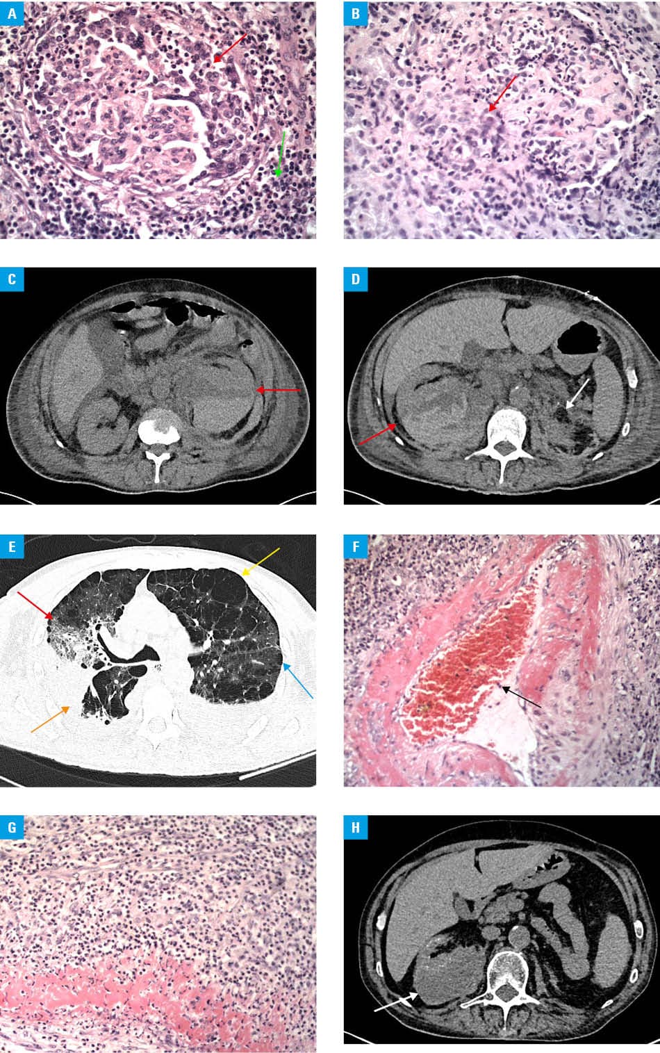
Three days after the biopsy, the patient presented with sharp abdominal pain and was found diaphoretic and hypotensive. Computed tomography (CT) showed massive subcapsular hematoma originating from the ruptured left kidney, penetrating to the retroperitoneal space and peritoneal cavity (Figure 1C). Immediate life‑saving nephrectomy was performed, with instant pathomorphological examination demonstrating multiple microabscesses. Due to this and persistent bacterial infection, we did not extend immunosuppression and continued GCSs with antibiotics, observing gradual improvement of the patient’s condition. However, 7 days after the nephrectomy, he experienced acute right‑sided abdominal pain and dyspnea. CT showed rupture of the right kidney with retroperitoneal hematoma infiltrating the renal capsule (Figure 1D) and new ground‑glass opacities within the lungs, suggesting diffuse alveolar hemorrhage (Figure 1E). Cyclophosphamide treatment was initiated, resulting in gradual improvement of the patient’s condition within the next 5 weeks. The patient did not require another laparotomy. Final microscopic evaluation of all specimens showed fibrinoid necrosis of medium‑sized arteries (Figure 1F) accompanying the aforementioned findings (Figure 1G), thus enabling a conclusive diagnosis of polyarteritis nodosa (PAN). With the continuation of GCSs and cyclophosphamide, the patient recovered and was found in a good condition 7 months later, although reliant on maintenance hemodialysis (Figure 1H).
While PAN can potentially affect any organ, very rarely does it cause glomerulopathy with the presence of ANCA antibodies in the serum.3 Whereas several cases of spontaneous kidney rupture in the course of PAN have been described,4,5 this might be the first documented case of bilateral kidney rupture. The presented case illustrates the complexity and heterogeneity of vasculitides.
- Baboudjian M, Lebacle C, Gondran‑Tellier B, et al. Who is at risk of death after renal trauma? An analysis of thirty‑day mortality after 1799 cases of renal trauma. Urol Int. 2023; 107: 165‑170. | Crossref
- Hellmich B, Sanchez‑Alamo B, Schirmer JH, et al. EULAR recommendations for the management of ANCA‑associated vasculitis: 2022 update. Ann Rheum Dis. 2024; 83: 30‑47. | Crossref
- Wolff L, Horisberger A, Moi L, et al. Polyarteritis nodosa: old disease, new etiologies. Int J Mol Sci. 2023; 24: 16668. | Crossref
- Kirkali Z, Finci R, Eryigit M, Ozdemir C. Spontaneous kidney rupture in polyarteritis nodosa. Report of four cases. Eur Urol. 1988; 15: 153‑155 | Crossref
- Banaszkiewicz M, Niezabitowska K, Krzanowski M, et al. Kidney rupture in a patient with polyarteritis nodosa. Pol Arch Intern Med. 2024; 134: 16759. | Crossref
SUPPLEMENTARY MATERIAL
ARTICLE INFORMATION