

 CC BY 4.0
Hypereosinophilic syndrome (HES) is defined by the presence of hypereosinophilia (HE) in the peripheral blood, tissue infiltration by eosinophils, and organ damage attributable to eosinophil‑mediated toxicity, provided that other underlying causes of organ dysfunction are excluded. HE is diagnosed when the absolute eosinophil count is equal to or greater than 1.5 × 10⁹/l on at least 2 occasions within a minimum interval of 2 weeks, or when tissue HE is demonstrated histologically. HES encompasses a heterogeneous group of disorders that may be classified as primary (clonal), secondary (reactive), or idiopathic, and its clinical spectrum ranges from indolent disease to life‑threatening organ complications, particularly involving the cardiovascular system.1,2
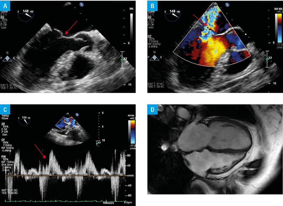
We present a case of a 63‑year‑old woman with long‑standing bronchial asthma and an episode of acute pulmonary embolism (APE) in the preceding month (the discharge recommendation was 20 mg of rivaroxaban once daily) admitted to a department of cardiology due to progressive dyspnea and 2 episodes of presyncope. On admission, she presented with New York Heart Association class III/IV symptoms, signs of pulmonary crackles, and a loud systolic murmur over the mitral valve. Transthoracic and transesophageal echocardiography showed severe mitral regurgitation (MR; Carpentier type II mechanism; rupture of the chordae tendineae and prolapse of A3 and A2 segments of the anterior mitral leaflet; Figure 1A–1C). Laboratory tests showed leukocytosis (leukocytes, 17.86 × 10³/µl; reference range [RR], 3.9–9.5 × 10³/µl]), marked eosinophilia (eosinophils, 8.26 × 10³/µl; RR, 0.03–0.39 × 10³/µl), and elevated concentrations of immunoglobulin E (4106 ng/ml; RR <240 ng/ml), N‑terminal pro–B‑type natriuretic peptide (433 pg/ml; RR, 0–125 pg/ml), and D‑dimer (2085.64 ng/ml; RR, 0–550 ng/ml). Retrospective analysis of APE hospitalization data did not demonstrate any abnormalities in complete blood count, eosinophil count, or mitral valve function. Among the differential diagnoses, a reactive form of HES potentially related to rivaroxaban was considered; therefore, the medication was discontinued and replaced with low‑molecular‑weight heparin. Oral prednisone therapy (20 mg daily) was initiated, resulting in rapid normalization of hematologic parameters. The acute MR required urgent qualification for cardiac surgery. Coronary angiography showed no significant coronary artery disease, and cardiac magnetic resonance (CMR) identified no specific abnormalities or features suggestive of eosinophilic myocardial infiltration (Figure 1D).The patient underwent successful surgical mitral valve repair, and no macroscopic signs of infiltration of the mitral valve structures were found intraoperatively. During multidisciplinary consultations, secondary causes of HE and clonal HE were excluded. The patient was diagnosed with seronegative eosinophilic granulomatosis with polyangiitis (EGPA; Churg–Strauss syndrome). The diagnosis was supported by the fulfilment of the 2022 American College of Rheumatology / European Alliance of Associations for Rheumatology classification criteria,3including long‑standing bronchial asthma and persistent peripheral blood eosinophilia (eosinophils ≥1000/µl), yielding a score equal to or higher than 6 points. Antineutrophil cytoplasmic antibodies negativity and the absence of hematuria or mononeuritis multiplex did not preclude the diagnosis. The summary of the diagnostic and therapeutic process is presented in Supplementary material. Long‑term treatment with oral corticosteroids and azathioprine was recommended, with consideration of biological therapy (mepolizumab) in the case of an inadequate response.
This case presents an atypical course of HES leading to a diagnosis of EGPA. Cardiac involvement in HES is common (reported in 60%–90% of the cases), and represents one of the leading causes of mortality. In this clinical case, acute MR occurred in the setting of HE and required urgent cardiac surgery. The key elements of qualification for the procedure were CMR imaging to assess the extent of eosinophilic infiltration, the presence of thrombi or myocardial fibrosis,4 and immediate and effective pharmacotherapy with steroids. A probable association between HE and the development of acute MR in the course of chordae tendineae rupture was considered, although definitive causality could not be established.
In the literature, only isolated case reports describe acute MR associated with papillary muscle rupture in patients with HES.5,6 This case highlights the necessity of a multidisciplinary approach to the diagnosis and management of patients with chronic HE of unclear etiology, and underscores the importance of a short‑term, decisive approach in situations involving urgent cardiac complications requiring surgical intervention.
- Valent P, Klion AD, Roufosse F, et al. Proposed refined diagnostic criteria and classification of eosinophil disorders and related syndromes. Allergy. 2023; 78: 47‑59. | Crossref
- Helbig G, Kyrcz‑Krzemień S. Diagnostic and therapeutic management in patients with hypereosinophilic syndromes. Pol Arch Med Wewn. 2011; 121: 44‑52. | Crossref
- Robson JC, Grayson PC, Ponte C, et al. 2022 American College of Rheumatology / European Alliance of Associations for Rheumatology classification criteria for granulomatosis with polyangiitis. Ann Rheum Dis. 2022; 81: 315‑320. | Crossref
- Michałowska I, Hryniewiecki T, Furmanek M. Imaging diagnostics. Heart and large vessels [in Polish]. Warszawa: PZWL; 2014. | Crossref
- Madhwal S, Goldberg J, Barcena J, et al. Unusual cause of acute mitral regurgitation: idiopathic hypereosinophilic syndrome. Ann Thorac Surg. 2012; 93: 974‑977. | Crossref
SUPPLEMENTARY MATERIAL
ARTICLE INFORMATION