
Pulmonary sarcoidosis initially suspected as post–COVID-19 syndrome: flow cytometry imaging of bronchoalveolar lavage

 CC BY 4.0
Pulmonary sarcoidosis initially suspected as post–COVID-19 syndrome: flow cytometry imaging of bronchoalveolar lavage
CC BY 4.0
A 46‑year‑old nonsmoking man, canary breeder, was admitted with suspected post–COVID‑19 syndrome (PCS) following a moderate SARS‑CoV‑2 infection 7 months earlier. Since that time, he had been experiencing persistent exertional dyspnea and fatigue.
Before contracting COVID‑19, the patient had no history of chronic disease or long‑term medication use. Routine occupational chest radiography had always been unremarkable. He reported no respiratory symptoms, even after substantial exposure to organic allergens during canary cage cleaning. He had received 2 doses of an mRNA COVID‑19 vaccine (Comirnaty; Pfizer‑BioNTech, New York, United States), the last administered 6 months before the acute infection.
Due to persistent symptoms, chest radiography was performed, showing peripheral band‑like and fine, patchy densities, predominately in the right lung, with hilar enlargement, interpreted as postinflammatory changes following SARS‑CoV‑2 infection (Figure 1A). Over the following weeks, the patient’s clinical improvement was only partial. Thirteen weeks after the acute phase of COVID‑19, he underwent pulmonary evaluation. His symptoms were initially attributed to PCS.
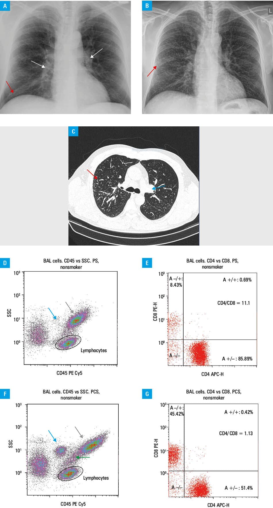
Abbreviations: APC‑H, hybrid argon plasma coagulation; BAL, bronchoalveolar lavage; PCS, post–COVID‑19 syndrome; PE‑H, phycoerythrin‑height; PE Cy5, phycoerythrin cyanine‑5; PS, pulmonary sarcoidosis; SSC, side scatter
Follow‑up chest radiography (Figure 1B) demonstrated abnormalities suggestive of interstitial lung disease (ILD) in the course of PCS; however, differential diagnosis with other forms of ILD remained necessary.
High‑resolution chest computed tomography performed 5 months after the acute illness showed bilateral, diffuse micronodular changes—mainly in the right upper lung lobe—accompanied by mediastinal lymphadenopathy (Figure 1C). Laboratory test results for SARS‑CoV‑2, influenza virus antigen assays, as well as precipitins to common hypersensitivity pneumonitis antigens and antinuclear antibodies were negative. Bacteriology was negative, as were acid‑fast bacilli smears and molecular testing for Mycobacterium tuberculosis complex DNA. Ultimately, the BACTEC blood culture system (Becton Dickinson Diagnostic Systems, Sparks, Maryland, United States) culture yielded no growth. Occupational history excluded exposure to inorganic dusts. Pulmonary function test results were within normal limits.
Endobronchial ultrasound identified enlarged mediastinal lymph nodes. Transbronchial needle aspiration of lymph node stations 4R and 7 showed aggregates of epithelioid cells, indicating the presence of granulomatous disease. Given the clinical presentation, sarcoidosis was considered.
Bronchoalveolar lavage (BAL) with lymphocyte immunotyping in flow cytometry demonstrated lymphocytic alveolitis (Figure 1D) and a very high CD4/CD8 index value of 11.1 (Figure 1E). Such a finding is typical of sarcoidosis,1 but not PCS. Of note, the CD4/CD8 index reference range for nonsmokers in our laboratory is 0.9–4.1
A final diagnosis of pulmonary sarcoidosis with mediastinal lymph node involvement (International Classification of Diseases, Tenth Revision code D86.2) was established, and an observational management strategy was adopted. At follow‑up, gradual regression of symptoms was observed.
PCS is common after acute COVID‑19, with pulmonary manifestations being particularly frequent.2 Sarcoidosis may develop following SARS‑CoV‑2 infection; however, an increase in its incidence in this population has not been conclusively proved—at most, the disease course may be more severe.3,4
Distinguishing PCS from sarcoidosis can be a significant clinical challenge. In contrast to the characteristic cytoimmunological profile in sarcoidosis, BAL in PCS typically demonstrates an increased total number of neutrophils and / or lymphocytes, a reduced CD4/CD8 index, and an increased neutrophil‑to‑lymphocyte ratio.5 Figure 1F and 1G illustrate distinct differences from sarcoidosis by showing analogous flow cytometric analysis of BAL cells in a PCS patient.
- Kopiński P, Wypasek E, Senderek T, et al. Different expression of immune checkpoint markers on bronchoalveolar lavage CD4+ cells: a comparison between hypersensitivity pneumonitis and sarcoidosis. Pol Arch Intern Med. 2021; 131: 16084. | Crossref
- Xie K, Zhang P, Li Y, Xia L. The post‑COVID‑19 pulmonary sequelae: manifestations, mechanisms and treatment strategies. J Thorac Dis. 2025; 17: 7414‑7429. | Crossref
- Bogdan M, Kanecki K, Tyszko P, et al. Hospitalizations of patients with sarcoidosis before and during the COVID‑19 pandemic in Poland. Pol Arch Intern Med. 2024; 134: 16618. | Crossref
- Lim SH, Ju HJ, Han JH, et al. Autoimmune and autoinflammatory connective tissue disorders following COVID‑19. JAMA Netw Open. 2023; 6: e2336120. | Crossref
- Dolna‑Michno J, Kopiński P, Przybylski G, et al. Cytoimmunological profile of lower airways in post‑COVID‑19 syndrome (PCS): predictive value of bronchoalveolar lavage. J Clin Med. 2025; 14: 3361. | Crossref
ARTICLE INFORMATION