


 CC BY-NC-SA 4.0
Time flies over us, but leaves its shadow behind.
Nathaniel Hawthorne
In cardiovascular medicine, as well as in health care at large, “time” is more than a stopwatch metric—it is in fact a biological coordinate shaping autonomic tone, endothelial function, coagulation balance, inflammatory response, and vulnerability to acute events.1,2 However, clock time and inner time can diverge, especially with an array of factors (eg, irregular sleep, shift work, jet lag, and chronic diseases) capable of disrupting healthy sleeping and, sometimes synergistically, leading to misalignment that matters both clinically and socially.3 Because patient symptoms, clinical decisions, and actual implementation steps also follow daily rhythms, the apparent circadian effects may reflect both physiology and the way patients and systems behave under pressure. Care for individuals experiencing acute atherothrombotic cardiovascular events, such as ST‑segment elevation myocardial infarction (STEMI), whose outcomes are unforgivingly time‑dependent, is therefore an ideal setting to test whether the inner clock leaves fingerprints on presentation and treatment.4 The nationwide registry analysis presented in this issue of Polish Archives of Internal Medicine is a timely contribution inviting us to ask not only when STEMI strikes, but also how timing influences recognition, reperfusion, and even the anatomy of culprit occlusion.5
Specifically, using the Polish National Percutaneous Coronary Intervention (PCI) Registry, Dziewierz et al5 reported on over 150 000 STEMI cases treated with primary PCI across more than 150 Polish centers in the years 2014–2022. Symptom onset showed a clear morning peak, followed by a smaller evening rise, reminding us that temporal clustering persists despite contemporary networks and pharmacotherapy. Crucially, the study links onset timing to the entire care pathway, separating patient delay from system delay by leveraging first medical contact and procedure time stamps. The authors also explored whether the infarct‑related artery differed by onset hour, an angle that shifts the discussion from “when” toward “where” in coronary anatomy. This combination of chronobiology and process analytics is indeed a highly useful contribution, because it identifies where the clock is a marker and where it might be a modifiable lever.
The most actionable finding was the nocturnal penalty in patient delay: symptoms beginning overnight were followed by substantially longer times to first medical contact than daytime events, with such a pattern likely reflecting sleep, symptom ambiguity, fear of overreacting, limited social support, and practical barriers to calling emergency services while still half‑awake. Importantly, the prolongation was amplified in older adults and in patients with diabetes, kidney disease, or chronic lung disease—groups already prone to atypical presentations. System delays also fluctuated, with longer intervals around the morning surge, suggesting that operational congestion and staffing rhythms can compound biological vulnerability at precisely the wrong moment. Seen through a quality lens, the nocturnal delay thus appears as a sanctionable prevention target, while the morning system delay represents more of a capacity‑planning problem that can be audited and improved.
The most hypothesis‑generating observation was an apparent interplay between the timing of STEMI and culprit lesion, with STEMIs due to left anterior descending artery (LAD) occlusion occurring proportionally more often at night, and those due to right coronary artery (RCA) occlusion being more common during daytime. If replicated, this finding would broaden the conversation from event timing to time‑dependent coronary physiology, including differences in phasic flow, shear stress, and plaque susceptibility between vessels. It also invites mechanistic thinking about sleep‑related stressors, such as intermittent hypoxemia and surges in sympathetic activity, which could significantly increase the risk in vulnerable patients overnight.6 However, anatomy is not sufficient per se as a causal factor, and several nonbiologic explanations must be considered, including differential symptom intensity, differential probability of awakening, and systematic recall error for nocturnal onset. A sensible clinical stance is to treat the LAD–RCA rhythm as a map for follow‑up studies, not as evidence that the clock selects a vessel in any deterministic way.
Methodologically, this work has several key strengths, including the national scale, representativeness, and granular, time‑stamped system intervals, appraised with appropriate effect estimates, while its principal limitation is reliance on patient‑reported symptom‑onset time.7 Nocturnal onset is especially vulnerable to rounding, uncertainty about when discomfort truly began, and misclassification between “woke with symptoms” and “symptoms started while asleep.”8 Future registries could capture awakening status, sleep interruption, and whether symptoms were recognized immediately or after a period of rest, improving sensitivity without jeopardizing specificity, as indeed hourly binning is intuitive, yet cyclic models could estimate phase and amplitude with confidence intervals, avoiding arbitrary cut points and enabling comparisons across regions and years. Notably, the caution showed by the authors in not performing too many exploratory analyses, particularly for periprocedural mortality, is welcome, because very large datasets can manufacture fragile signals that vanish after adjustment.
For patients, the practical message is simple and urgent: if chest discomfort, unexplained dyspnea, diaphoresis, or arm or jaw discomfort wakes you from sleep, call emergency services immediately (Figure 1). If symptoms begin at night, do not wait until daytime, as the registry shows that night‑time events are exactly when help‑seeking delays are longest and salvageable myocardium is lost.9 Wearables and connected sensors can complement this message by detecting sustained tachycardia, rhythm irregularity, nocturnal hypoxemia, or abrupt activity changes, and by time‑stamping symptom‑linked physiological shifts.10 When integrated into internet of things–enabled care pathways, these streams can trigger earlier tele‑triage, transmit single‑lead electrocardiograms (ECGs), and shorten the path from suspicion to activation. Artificial intelligence models, if carefully validated, may also help distinguish benign noise from high‑risk patterns, personalize alerts for high‑risk patients, and provide operation teams with demand forecasts aligned to circadian peaks. For physicians, the paper argues for making “time” a part of the clinical phenotype, documenting symptom‑onset context, sleep disruption, and possible sleep apnea in high‑risk patients. For health care managers, the registry suggests a new form of quality improvement: dashboard pain‑to‑contact and contact‑to‑device times by hour, stratify by vulnerability, and intervene where gaps predictably cluster.11 Morning surges imply the need for capacity planning and rapid activation pathways, whereas nocturnal delays point toward community education, dispatch readiness, and messaging that legitimizes calling at night. Operational fixes may include prehospital ECG routing, streamlined catheterization laboratory activation protocols, and staffing models that match peak demand without compromising off‑hours safety and supervision. Hospitals should also recognize their own circadian footprint, minimizing unnecessary nocturnal sleep disruption when safe, because fragmented sleep may worsen autonomic stress and recovery in cardiac patients.
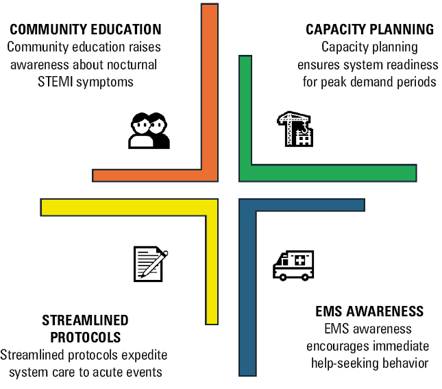
Abbreviations: EMS, Emergency Medical Services
In conclusion, the clock is not fate, but it is timely information. If used wisely, it can guide mechanistic discovery, sharpen prevention, and make STEMI systems faster and fairer at every hour.
- Chellappa SL, Vujovic N, Williams JS, Scheer FAJL. Impact of circadian disruption on cardiovascular function and disease. Trends Endocrinol Metab. 2019; 30: 767‑779. | Crossref
- Biondi‑Zoccai GG, Abbate A, Liuzzo G, Biasucci LM. Atherothrombosis, inflammation, and diabetes. J Am Coll Cardiol. 2003; 41: 1071‑1077. | Crossref
- Paschos GK, FitzGerald GA. Circadian clocks and vascular function. Circ Res. 2010; 106: 833‑841. | Crossref
- Del Prete A, Conway F, Della Rocca DG, et al. COVID‑19, acute myocardial injury, and infarction. Card Electrophysiol Clin. 2022; 14: 29‑39. | Crossref
- Dziewierz A, Malinowski K, Dardzińska N, et al. Circadian variation in ST‑segment elevation myocardial infarction: a nationwide analysis of onset, treatment delays, and culprit artery patterns. Pol Arch Intern Med. 2026; 136: 17188. | Crossref
ARTICLE INFORMATION