

 CC BY 4.0
A 24‑year‑old woman was admitted after a pulmonary lesion was incidentally detected during a routine health check‑up. She was asymptomatic, and both physical examination and laboratory test results were unremarkable. Contrast‑enhanced chest computed tomography (CT) identified a heterogeneous mass measuring 11.3 cm × 8.1 cm in the right lower lobe, containing gas, soft‑tissue, as well as calcified and fat‑density components, with delayed enhancement of soft‑tissue elements (Figure 1A–1D).
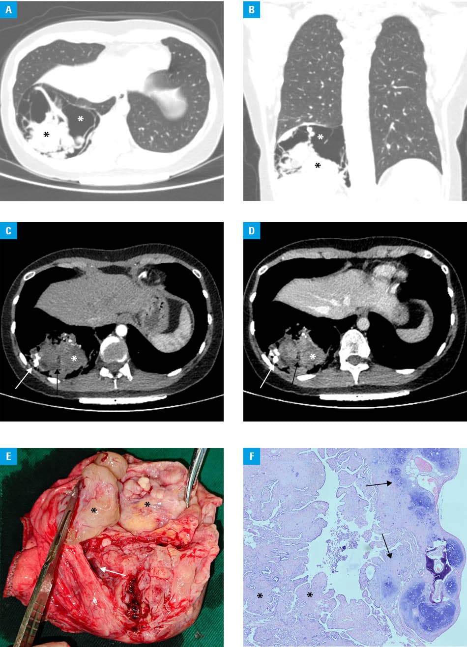
The patient underwent video‑assisted thoracoscopic surgery under general anesthesia, during which the mass, along with a small margin of adjacent healthy lung parenchyma, was resected using a surgical stapler. The resected specimen weighed 180 g. A well‑defined mass adjacent to the pleura was identified. The cut surface had a mixed cystic and solid appearance, with multiple smooth‑walled cysts ranging from 1 to 5 cm in diameter. The solid component accounted for approximately 80% of the lesion, it was firm and brown, and had areas of bony hardness and focal yellowish discoloration (Figure 1E). Microscopic examination demonstrated numerous cystic spaces lined with the respiratory epithelium. The solid stromal component was composed predominantly of fibrous tissue and contained smooth muscle, cartilage, bone, and areas of calcification (Figure 1F). A final diagnosis of adenofibromatous hamartoma with scarce cartilage was established. The patient recovered uneventfully and was discharged on postoperative day 3.
Hamartomas are characterized by a disorganized mixture of epithelial and mesenchymal tissues. Pulmonary hamartoma (PH) is a common benign tumor that is most frequently diagnosed in adults during the fifth to sixth decades of life. PHs are typically solitary, peripherally located, and well‑circumscribed lesions, usually measuring 1–4 cm in diameter (average, 2 cm).1 Larger lesions or multiple hamartomas have been reported occasionally.2,3 PHs are categorized according to their dominant component into chondroid, lipomatous, adenoleiomyomatous, and adenofibromatous subtypes, with the chondroid subtype being the most prevalent. The presence of “popcorn” calcification and fat density within the lesion supports a diagnosis of PH.1 The multicystic, heterogeneous mass in this patient differed from the typical, giant, or adenofibromatous hamartomas previously reported in the literature.2,3
The primary differential diagnosis in the case of our patient included mediastinal teratoma with rupture into the lung and congenital pulmonary airway malformation (CPAM). Teratomas typically appear as complex masses composed of soft tissue, calcification, fat, and fluid. However, in the absence of rupture into the lung, a gas component is typically not observed. Moreover, intrapulmonary teratomas are exceedingly rare, as compared with mediastinal teratomas. In this patient, the lesion was centered within the lung, well‑circumscribed, and contained gas, which was not consistent with a teratoma. CPAM may appear as a heterogeneous lesion containing varying proportions of fluid and gas, and is most commonly diagnosed in infants or fetuses. However, CPAM typically lacks calcification and fat, which were present in this patient.
This case of a multicystic 11.3‑cm PH in a 24‑year‑old woman is remarkable due to the patient’s young age, large tumor size, and atypical imaging features.It expands the clinical understanding of PH, and may inform a more individualized treatment approach.
- Siegelman S, Khouri NF, Scott Jr W, et al. Pulmonary hamartoma: CT findings. Radiology. 1986; 160: 313‑317. | Crossref
- Ozbudak IH, Dertsiz L, Bassorgun CI, Ozbilim G. Giant cystic chondroid hamartoma of the lung. J Pediatr Surg. 2008; 43: 1909‑1911. | Crossref
- Saadi MM, Barakeh DH, Husain S, Hajjar WM. Large multicystic pulmonary chondroid hamartoma in a child presenting as pneumothorax. Saudi Med J. 2015; 36: 487‑489. | Crossref
ARTICLE INFORMATION