

 CC BY 4.0
Hemophagocytic lymphohistiocytosis (HLH) is a rare, life‑threatening, and often fatal, cytokine‑mediated, hyperinflammatory syndrome. It can be challenging to diagnose because the symptoms are nonspecific and resemble those of other, more common conditions, such as sepsis.1,2 Primary HLH is associated with underlying genetic mutations, and is often diagnosed in children. Conversely, secondary HLH usually occurs in adults, and is most often triggered by infection, malignancy, or autoimmune disorders.1,2 Its exact pathomechanism remains poorly understood, but it is known to result from uncontrolled and persistent activation of cytotoxic T cells and natural killer cells.1,3 Defining symptoms are listed in the HLH‑2004 criteria2. The incidence of secondary HLH remains unclear and is most likely severely underestimated due to the diagnostic challenges posed by the disease.2-4
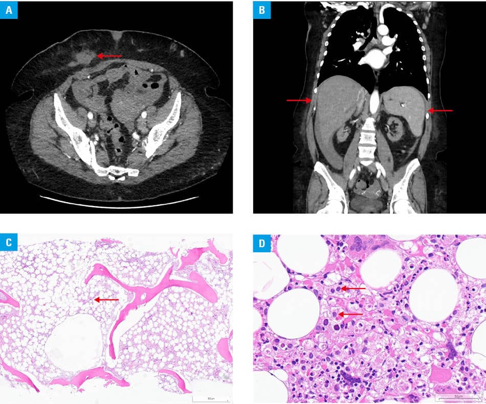
We present the case of a 46‑year‑old woman with end‑stage chronic kidney disease of unknown etiology. She underwent peritoneal dialysis for 4 years without complications and was admitted to a hospital with a suspected thrombus in her left lower limb veins. Although the diagnosis of thrombosis was excluded, she was urgently admitted to a nephrology department due to bleeding disorders, elevated inflammatory markers (C‑reactive protein, 93.4 mg/l; reference range [RR] <5 mg/l), and severe anemia. Activated partial thromboplastin time and international normalized ratio were prolonged. She received empirical antibiotic, antiviral, and antifungal therapies, as well as blood product transfusions. The patient was converted to hemodialysis due to peritoneal dysfunction, as well as high serum creatinine and urea concentrations. Numerous microbiological cultures and virological test results were negative, including hepatitis B surface antigen, hepatitis B surface antibody, and hepatitis C surface antibody, as well as polymerase chain reaction tests for HIV, parvovirus B19, cytomegalovirus, Epstein–Barr virus, SARS‑CoV‑2, influenza A and B (including subtypes), 4 types of coronaviruses, 4 types of parainfluenzas, respiratory syncytial virus, human metapneumovirus, adenovirus, rhinovirus, enterovirus, and rotavirus. Transthoracic and transesophageal echocardiography, bronchoscopy, exploratory laparoscopy, and contrast‑enhanced computed tomography (Figure 1A and 1B) were performed to identify the source of infection, but they were unremarkable. The patient subsequently developed episodes of persistent fever, thrombocytopenia (platelets, 86 × 10³/μl; RR, 140–440 × 10³/μl), and had elevated levels of serum ferritin (172 000 μg/l; RR, 30–400 μg/l) and interleukin 6 (104 pg/ml; RR <7 pg/ml). She also experienced leukopenia (leukocytes, 3.8 × 10³/μl; RR, 4–10 × 10³/μl) and neutropenia (neutrophils, 0.9 × 10³/μl; RR, 1.8–7.72 × 10³/μl). HLH was suspected, so aspiration biopsy and bone marrow trephine biopsy were performed (Figure 1C and 1D). The H‑score of the patient was 284, indicating an over 99% probability of this diagnosis. Treatment according to the HLH protocol was initiated prior to receiving histopathology results, and included dexamethasone, etoposide, and intravenous immunoglobulins. Despite treatment according to the established recommendations, the patient died.
This case underscores the diagnostic challenges associated with secondary HLH, particularly among patients undergoing peritoneal dialysis. Although several cases of this complication have been reported in the literature, most are linked to infections, which are a common trigger of secondary HLH. Recently, however, research has suggested that chronic exposure to peritoneal dialysis fluids provokes local cellular and molecular effects, including sterile inflammation. These effects not only cause local lesions, but also trigger systemic metabolic, inflammatory, and immune‑modulatory effects.5 HLH should be considered in the cases of unexplained, persistent systemic inflammation with multiorgan dysfunction, cytopenias, and elevated inflammatory markers, particularly hyperferritinemia. Although standardized treatment protocols improve mortality rates in HLH patients, they still remain high. Thus, early diagnosis and appropriate treatment are crucial for patient survival.
- La Rosée P, Horne AC, Hines M, et al. Recommendations for the management of hemophagocytic lymphohistiocytosis in adults. Blood. 2019; 133: 2465‑2477. | Crossref
- Konkol S, Killeen MB, Rai M. Hemophagocytic lymphohistiocytosis. In: StatPearls. Treasure Island (FL): StatPearls Publishing; 2025 May. https://www.ncbi.nlm.nih.gov/books/NBK557776/. Accessed May 3, 2025. | Crossref
- Sepulveda FE, de Saint Basile G. Hemophagocytic syndrome: primary forms and predisposing conditions. Curr Opin Immunol. 2017; 49: 20‑26. | Crossref
- Ponnatt TS, Lilley CM, Mirza KM. Hemophagocytic lymphohistiocytosis. Arch Pathol Lab Med. 2022; 146: 507‑519. | Crossref
- Trionfetti F, Marchant,V, Guadalupe T, et al. Novel aspects of the immune response involved in the peritoneal damage in chronic kidney disease patients under dialysis. Int J Mol Sci. 2023; 24: 5763. | Crossref
ARTICLE INFORMATION