
Two in one: paraganglioma and gastrointestinal stromal tumor presentation of Carney–Stratakis syndrome

 CC BY 4.0
Two in one: paraganglioma and gastrointestinal stromal tumor presentation of Carney–Stratakis syndrome
CC BY 4.0
A 31‑year‑old patient with no previous medical history presented to a hospital with gastrointestinal bleeding. Laboratory test results showed anemia (hemoglobin, 10 g/dl; reference range, 14–18 g/dl). Diagnostic workup, including gastroduodenoscopy, identified a submucosal tumor with ulceration in the stomach body, while abdominal computed tomography (CT) demonstrated gastric and paraaortic lymphadenopathy.
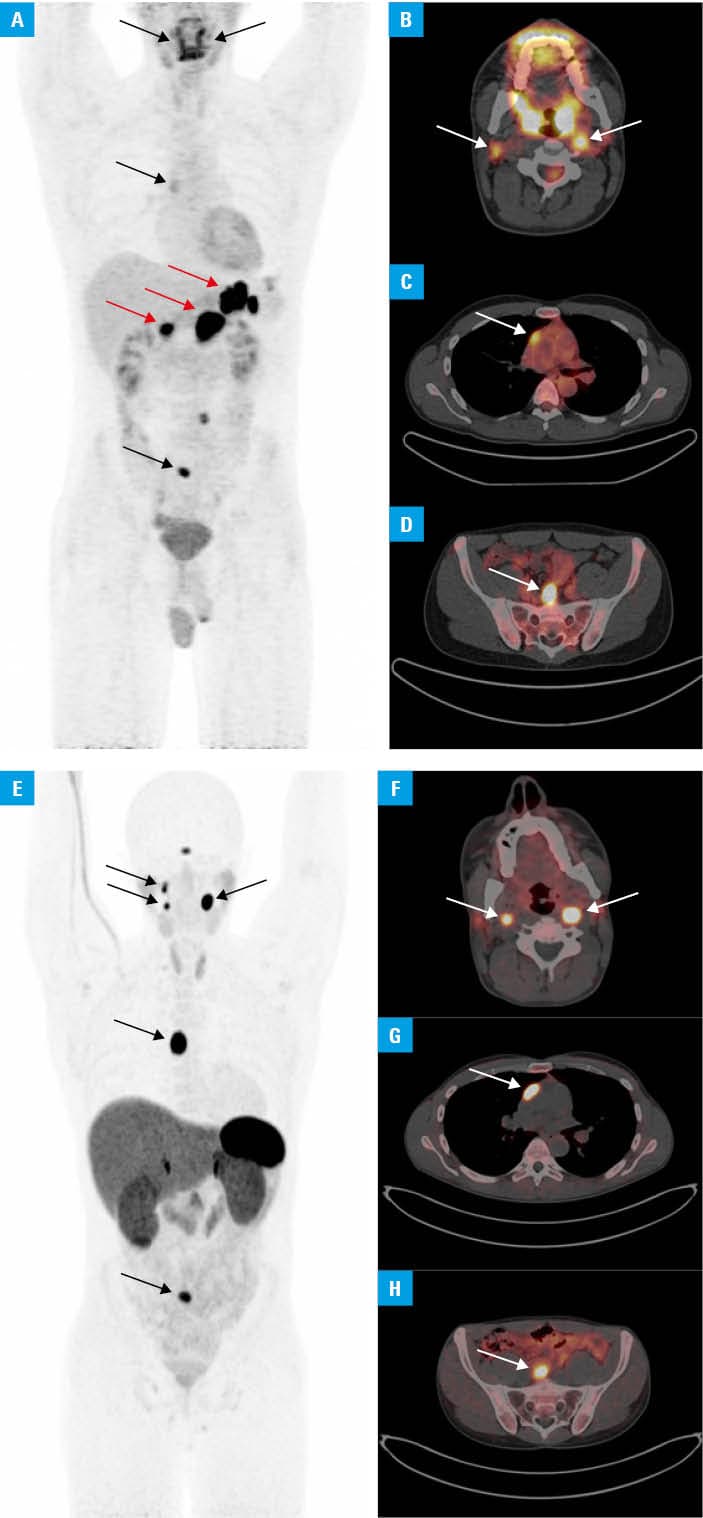
18F‑fluorodeoxyglucose positron emission tomography / computed tomography (18F‑FDG PET/CT) showed multiple lesions (Figure 1A) with increased focal radiotracer uptake in the gastric wall, enlarged para‑aortic and perigastric lymph nodes (LNs), as well as additional foci bilaterally near the jugular vein openings (Figure 1B), adjacent to the aortic bulb (Figure 1C), and in the presacral region (interpreted as a metastatic LN; Figure 1D). Histopathological examination of the endoscopic biopsy confirmed a gastrointestinal stromal tumor.
Oncologists initiated imatinib1 chemotherapy. After 6 months, the patient underwent subtotal gastrectomy with gastrointestinal reconstruction; para‑aortic and gastric LNs were also resected. Surprisingly, histopathology identified 1 LN as paraganglioma rather than gastrointestinal stromal tumor (GIST) metastasis.
Follow‑up 18F‑FDG PET/CT showed a stable metabolically active lesion in the sacral area, which could represent either GIST metastasis or another paraganglioma. Given the patient’s young age, good clinical condition, and the need for accurate diagnosis to guide management, somatostatin receptor imaging with gallium‑68 DOTA‑D‑Phe1‑Tyr3‑octreotate PET/CT was performed (Figure 1E). This showed multifocal radiotracer uptake, including in the previously 18F‑FDG–avid lesions (except the surgical lodge), confirming multifocal paraganglioma (Figure 1F–1H).2
These clinical and imaging findings raised a suspicion of Carney–Stratakis syndrome, a rare condition featuring both paraganglioma and GIST—distinct from Carney triad,3 which additionally includes pulmonary chondromas. With no lesion growth or their hormonal activity, the patient remains under active surveillance. Genetic testing was performed, ruling out the most common SDH mutations. Due to the possibility of other, rarer genetic mutations, the patient was referred for more detailed genetic testing,4 which is currently underway.
- Serrano C, Martín‑Broto J, Asencio‑Pascual JM, et al. 2023 GEIS Guidelines for gastrointestinal stromal tumors. Ther Adv Med Oncol. 2023; 15: 17588359231192388. | Crossref
- Zhi Y, Gerhard‑Hartmann E, Hartrampf PE, et al. Somatostatin receptor‑directed PET/CT can differentiate between different subtypes of head and neck paragangliomas. Clin Nucl Med. 2023; 48: 923‑927. | Crossref
- Stratakis CA, Carney JA. The triad of paragangliomas, gastric stromal tumours and pulmonary chondromas (Carney triad), and the dyad of paragangliomas and gastric stromal sarcomas (Carney–Stratakis syndrome): molecular genetics and clinical implications. J Intern Med. 2009; 266: 43‑52. | Crossref
- Freitas‑Castro F, Santana LS, Fagundes GFC, et al. SLC25A11, a novel gene associated with Carney–Stratakis syndrome. J Endocr Soc. 2025; 9: bvaf052. | Crossref
ARTICLE INFORMATION