

 CC BY 4.0
Danon disease (DD) is a rare X‑linked lysosomal myopathy caused by mutations in the LAMP2 gene.1 In women, the clinical phenotype usually manifests later than in men, typically in the third decade of life,2 ranging from isolated hypertrophic cardiomyopathy (HCM) to progressive heart failure and complex arrhythmias.3,4 We present 2 unrelated cases of early‑onset DD, illustrating its complex clinical course in women. Both presented in childhood with an isolated cardiac phenotype and electrocardiography (ECG) features of pre‑excitation (Figure 1A). A 3‑generation pedigree analysis showed no family history of cardiomyopathy, skeletal myopathy, or sudden cardiac death.
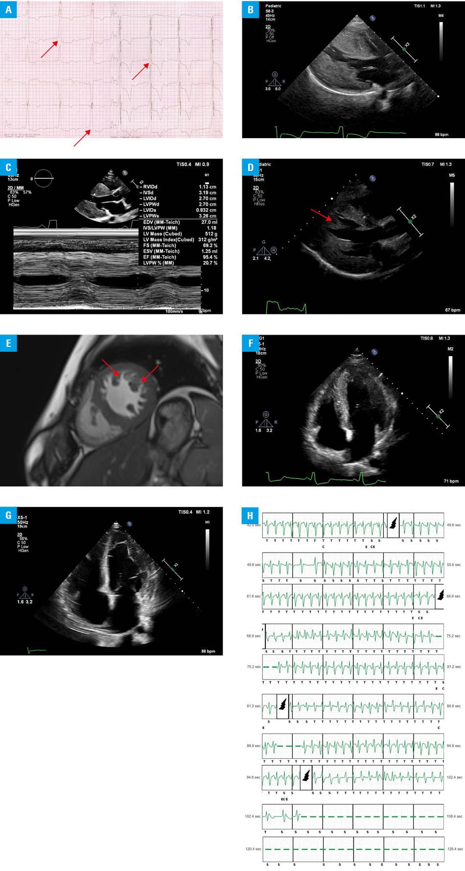
Abbreviations: EDV, end‑diastolic volume; EF, ejection fraction; ESV, end‑systolic volume; FS, fractional shortening; HGen, harmonic general; IVS, interventricular septum; LVID, left ventricular internal diameter; LVPW, left ventricular posterior wall; MI, mechanical index; MM, motion mode; RVID, right ventricular internal diameter
Patient 1 presented at the age of 11 years with exertional dyspnea persisting for 3 months. Physical examination showed a systolic murmur (Levine scale, 3/6). Laboratory test results demonstrated markedly elevated levels of high‑sensitivity troponin I (1923 ng/l; reference range [RR] <19 ng/l) and N‑terminal pro–B‑type natriuretic peptide (NT‑proBNP; 7740 pg/ml; RR <125 pg/ml). Transthoracic echocardiography (TTE) identified extreme left ventricular hypertrophy (LVH; 18.3 mm; Z‑score, +13) with diastolic dysfunction and elevated LV ejection fraction (LVEF 88%; Figure 1B). Cardiac magnetic resonance (CMR) showed intramural fibrosis. Genetic testing identified a pathogenic nonsense mutation in the LAMP2 gene (NM_002294.3:c.329C>A, p.Ser110*). The mutation was absent in both parents, confirming its de novo origin. Furthermore, a variant of uncertain significance in the MYBPC3 gene (NM_000256.3:c.49C>T, p.Arg17Trp) was identified. Segregation analysis showed maternal inheritance and presence in the asymptomatic sister, supporting the LAMP2 gene mutation as the primary pathogenic variant, although a modifier role of the MYBPC3 gene could not be excluded.
Over 3 years, LVH progressed markedly (Figure 1C). Given nonsustained ventricular tachycardia (nsVT) on Holter ECG and rapid disease progression, a subcutaneous implantable cardioverter‑defibrillator (S‑ICD) was implanted. At the of age 18 years, the patient remains stable (New York Heart Association class II) with preserved systolic function.
Patient 2 presented at the age of 14 years with a 6‑month history of palpitations, chest pain, and exertional dyspnea. Laboratory test results showed elevated levels of high‑sensitivity troponin I (353 ng/l) and NT‑proBNP (485 pg/ml). TTE identified asymmetric hypertrophy of the interventricular septum, increased LVEF, and LV mass (Figure 1D). CMR demonstrated mixed phenotype of HCM and LV noncompaction (Figure 1E). Electrophysiological study (EPS) confirmed a right‑sided accessory atrioventricular pathway, successfully treated with radiofrequency ablation, although pre‑excitation persisted thereafter. Genetic analysis identified a heterozygous deletion in the LAMP2 gene (NM_002294.3:c.451–?923+?del). The finding was verified by array comparative genomic hybridization (arr[GRCh37] Xq24[119580227 × 2, 119582863_119582922 × 1, 119584302 × 2]) and targeted real‑time polymerase chain reaction testing (rsa Xq24[LAMP2exons4‑5] × 1), showing a deletion of exons 4 and 5 (<4075 base pairs). Parental screening confirmed de novo status of the mutation.
The clinical course was dominated by progressive myocardial fibrosis on CMR and increasing arrhythmic burden. At the age of 17 years, given episodes of nsVT, an S‑ICD was implanted. Within 24 months, the patient underwent a phenotypic transition from hypertrophic to dilated cardiomyopathy (Figure 1F and 1G). The progression was complicated by LV thrombus, electrical instability with multiple inappropriate S‑ICD shocks (Figure 1H), and cardiogenic shock. The patient ultimately underwent heart transplantation at the age of 20 years, complicated by early graft failure (requiring venoarterial extracorporeal membrane oxygenation) and acute kidney injury. At 2‑year follow‑up, the patient remains stable.
Early‑onset DD in women may present as severe cardiac hypertrophy and malignant arrhythmias, mimicking the more aggressive male phenotype and even leading to rapid progression into dilated phenotype. These observations highlight the importance of vigilant monitoring for high‑risk markers, including hypertrophy progression and nsVT, particularly in the absence of validated risk stratification tools. Patient 1 lacked typical symptoms of arrhythmia despite extreme hypertrophy and documented nsVT; therefore, EPS was deferred. Notably, EPS performed in patient 2 due to palpitations before genetic diagnosis demonstrated a rare dual mechanism: a true accessory pathway amenable to ablation, coexisting with pseudo–pre‑excitation typical of DD, supporting electrophysiological evaluation in symptomatic pre‑excitation.
- Nishino I, Fu J, Tanji K, et al. Primary LAMP‑2 deficiency causes X‑linked vacuolar cardiomyopathy and myopathy (Danon disease). Nature. 2000; 406: 906‑910. | Crossref
- Boucek D, Jirikowic J, Taylor M. Natural history of Danon disease. Genet Med. 2011; 13: 563‑568. | Crossref
- Brambatti M, Caspi O, Maolo A, et al. Danon disease: gender differences in presentation and outcomes. Int J Cardiol. 2019; 286: 92‑98. | Crossref
- Hong KN, Eshraghian E, Khedro T, et al. An international longitudinal natural history study of patients with Danon disease: unique cardiac trajectories identified based on sex and heart failure outcomes. J Am Heart Assoc. 2025; 14: e038394. | Crossref
ARTICLE INFORMATION