
First report of enchondroma mimicking thyroid cancer metastasis on post-therapeutic 131I scintigraphy and single photon emission computed tomography / computed tomography
2 ,
,

 CC BY 4.0
First report of enchondroma mimicking thyroid cancer metastasis on post-therapeutic 131I scintigraphy and single photon emission computed tomography / computed tomography
, CC BY 4.0
A 32‑year‑old woman presented to an endocrinology department for radioactive iodine (RAI) adjuvant therapy for thyroid carcinoma. The patient’s medical history included kidney transplantation a year earlier due to end‑stage renal failure caused by recurrent severe nephrolithiasis. Three months before admission, she underwent total thyroidectomy with central neck dissection, and was diagnosed with papillary thyroid carcinoma (PTC), classic subtype pT1b N1a (ENE–) LVI1 Rx according to the 2017 American Joint Committee on Cancer classification, with an intermediate‑to‑high risk of recurrence according to the 2025 American Thyroid Association (ATA) guidelines.1 Genetic testing of the tumor tissue identified a somatic BRAF V600E pathogenic variant.
On admission, bilateral thyroid remnants were found on neck ultrasound. The thyroglobulin concentration was 0.71 μg/l (reference range [RR] for excellent response to surgical‑only treatment according to the 2025 ATA guidelines is <2.5 μg/l),1 the thyroid stimulating hormone (TSH) level was 0.624 mIU/l (RR, 0.1–0.5 mIU/l), the antithyroglobulin antibody level was 16.5 kIU/l (RR <115 kIU/l), and the stimulated thyroglobulin concentration was 2.43 μg/l. The patient received 2091 MBq (56.5 mCi) of RAI following exogenous recombinant TSH stimulation. On post‑therapeutic whole‑body scintigraphy (WBS), focal RAI uptake in the right shoulder was visualized (Figure 1A). Single photon emission computed tomography / computed tomography (SPECT/CT) of the neck and chest showed RAI uptake area in the right humeral head (Figure 1B), without evidence of structural bone remodeling on CT (Figure 1C). The cortical bone layer was preserved. No other foci of pathological RAI uptake were detected. On magnetic resonance imaging (MRI), a focal lesion measuring 2 mm × 7 mm × 6 mm in the humeral head, at the level of the lesser tuberosity, was observed. It was hypointense on T1‑weighted images (Figure 1D) and hyperintense on T2‑weighted, short τ inversion recovery, and proton density fat‑saturated sequences (Figure 1E), with no diffusion restriction. There was a rim of peripheral contrast enhancement (Figure 1F) with chemical shift artifacts present in the peripheral portions on T1‑weighted images (Figure 1G). The morphology and signal characteristics suggested a lesion consistent with an enchondroma, a benign cartilaginous tumor. The patient was referred to a department of orthopedic surgery for further management.
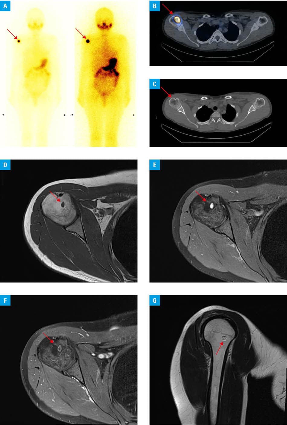
To our knowledge, this is the first case in the literature to report RAI uptake in an enchondroma. Case reports and case series of false‑positive RAI uptake in benign lesions have been published previously, most commonly describing inflammatory or benign lesions of various origins. The occurrence of these findings is estimated to be around 1%, potentially diminishing the utility of that imaging in low‑risk cases.2 The causes of false‑positive imaging involve contamination, inflammation, and passive accumulation.3 However, the mechanism of the RAI uptake observed within an enchondroma is unknown, and any explanation remains speculative. Enchondromas are hypocellular lesions, and chondrocytes are not known to express the sodium / iodide symporter. Diffusion to the extracellular portion of the lesion, with reduced clearance and gradual accumulation of RAI, seems therefore more probable. SPECT/CT is the imaging modality of choice when RAI pathological uptake is identified on WBS, as it allows for spatial recognition and better characterization of the uptake focus.3 Other imaging modalities (such as MRI) should be employed for further investigation in uncertain cases. A low thyroglobulin concentration, consistent with an excellent response to surgical‑only treatment, was another argument supporting the low probability of distant metastases in our patient. Furthermore, distant metastases to the bones are rare and occur more frequently in follicular and oncocytic thyroid cancers that spread hematogenously than in PTC, in which lymphatic spread and lung metastases are more common.4,5 Thus, the clinical context and assessment of cancer biomarkers should always be taken into account to avoid overtreatment.
- Ringel MD, Sosa JA, Baloch Z, et al. 2025 American Thyroid Association Management Guidelines for Adult Patients with Differentiated Thyroid Cancer. Thyroid. 2025; 35: 841‑985. | Crossref
- Oral A, Yazıcı B, Eraslan C, Burak Z. Unexpected false‑positive I‑131 uptake in patients with differentiated thyroid carcinoma. Mol Imaging Radionucl Ther. 2018; 27: 99‑106. | Crossref
- Barbaro D, Campennì A, Forleo R, Lapi P. False‑positive radioiodine uptake after radioiodine treatment in differentiated thyroid cancer. Endocrine. 2023; 81: 30‑35. | Crossref
- Iñiguez‑Ariza NM, Bible KC, Clarke BL. Bone metastases in thyroid cancer. J Bone Oncol. 2020; 21: 100282. | Crossref
- Boucai L, Zafereo M, Cabanillas ME. Thyroid cancer: a review. JAMA. 2024; 331: 425‑435. | Crossref
ARTICLE INFORMATION