
Unusual cause of severe abdominal pain in a young woman: concurrent nutcracker syndrome and median arcuate ligament syndrome

 CC BY 4.0
Unusual cause of severe abdominal pain in a young woman: concurrent nutcracker syndrome and median arcuate ligament syndrome
CC BY 4.0
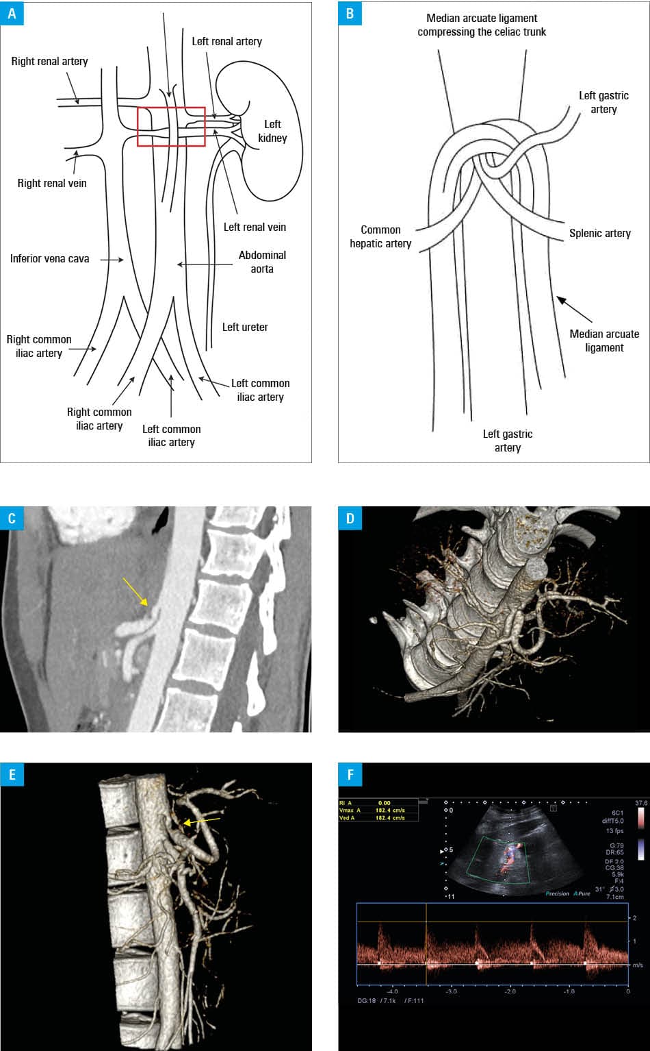
Median arcuate ligament syndrome (MALS; also referred to as Dunbar syndrome) and nutcracker syndrome (NCS) are rare vascular compression disorders presenting with nonspecific abdominal pain (Figure 1A and 1B).1 Their coexistence is extremely rare and complicates diagnostic evaluation.2,3
A 33‑year‑old woman with unremarkable medical history was admitted to a hospital due to recurrent severe, diffuse abdominal pain persisting for 4 days, radiating to the left lumbar region and worsening with movement. The pain initially occurred approximately 1 week prior to the current hospitalization. Contrast‑enhanced computed tomography of the abdomen and pelvis performed during previous hospitalization in a regional hospital raised suspicion of NCS. On admission, the patient was alert, hemodynamically stable, and reported severe abdominal pain rated up to 9 out of 10 points on the Numeric Rating Scale (NRS). The pain was unrelated to food intake and worsened when the patient moved or assumed an upright position, with significant improvement while lying down and with knees drawn up toward the chest. Abdominal ultrasonography and radiography of the chest and abdomen were unremarkable. Laboratory parameters, including complete blood count, serum biochemistry, inflammatory markers, and urinalysis, were all within reference ranges. Abdominal examination showed tenderness in the lower quadrants without guarding, rebound tenderness, or palpable masses. To exclude alternative etiologies, magnetic resonance imaging of the lumbosacral spine was performed, showing only mild L5–S1 discopathy without nerve root compression, unrelated to the symptoms.
Despite escalating analgesic therapy, including nonsteroidal anti‑inflammatory drugs and spasmolytics, the patient continued to experience pain at the level of 6–9 points on the NRS. Only morphine provided temporary relief. Renal Doppler ultrasonography confirmed significant compression of the left renal vein between the aorta and the superior mesenteric artery (SMA), consistent with anterior NCS (SMA peak systolic velocity [PSV], 170 cm/s; normal range 97–142 cm/s). Additionally, Doppler ultrasound evaluation of the celiac axis demonstrated a patent but markedly narrowed proximal celiac trunk (PSV, 260 cm/s; normal range 98–105 cm/s) due to extrinsic compression by the median arcuate ligament of the diaphragm. The combination of imaging findings (Figure 1C–1F) and clinical presentation supported the diagnosis of concurrent NCS and MALS.
Throughout hospitalization, the patient continued to experience severe abdominal pain, particularly nocturnal, with temporary improvement only after opioid administration. Over the following days, her symptoms gradually subsided, and she was discharged in a stable condition with a referral to a vascular surgery department at our hospital for further management.
About a month later, the patient was readmitted due to another episode of severe abdominal pain, and was referred for a surgical evaluation. She was subsequently qualified for open median arcuate ligament release surgery, as MALS seemed to be the most possible cause of her clinical condition. After successful surgery, pain resolved completely; the patient was discharged and no pain recurrence was observed during follow‑up.
Previous reports of concurrent NCS and MALS comprise a case of NCS and MALS with superadded gastroparesis and bradygastria,2 as well as a case of MALS contributing to the development of varicocele and NCS.4 A recent review outlined the diagnosis and treatment of NCS, but not its association with MALS.5 It should be stressed that the overlap between these disorders and the potential for multiple coexistent conditions which complicate diagnosis can cause increased lead time and morbidity for patients.
- Cardoso DL, De Macedo BA, Neto RM, et al. Abdominal vascular compression syndromes: a pictorial review. Eur J Radiol. 2025; 189: 112169. | Crossref
- Donnelly L, Turner B, Davies AH. Atypical case of coexistent vascular compression syndromes: median arcuate ligament syndrome and nutcracker syndrome. BMJ Case Rep. 2023; 16: e257754. | Crossref
- Moreno MC, Galván F, Cortés SX, Caunedo ÁÁ. An unusual association: median arcuate ligament syndrome and nutcracker syndrome. Rev Esp Enferm Dig. 2019; 111: 241‑242. | Crossref
- Shah S, Patel N, Spitzer L. Median arcuate ligament syndrome causing Nutcracker syndrome and left‑sided varicocele. Cureus. 2025; 17: e83177. | Crossref
- Mačionienė E, Kerpauskienė A, Žakauskienė U, et al. Nutcracker syndrome in 2026: a nephrologist‑oriented diagnosis and management. Clin Kidney J. 2026; 19: sfag124. | Crossref
ARTICLE INFORMATION