


 CC BY-NC-SA 4.0
A 64‑year‑old woman with genetically confirmed Fabry disease caused by a pathogenic GLA variant (c.644A>G; p.Asn215Ser), consistent with a late‑onset phenotype, was referred for evaluation of exertional chest pain (Canadian Cardiovascular Society class II). Her comorbidities included arterial hypertension and dyslipidemia, both well‑recognized cardiovascular risk factors. Plasma lyso‑Gb3 level was 3.1 ng/ml (reference range [RR] <1.8 ng/ml). The patient had been receiving enzyme replacement therapy with agalsidase β at a dose of 1 mg/kg, administered intravenously every other week for the past year. Transthoracic echocardiography demonstrated preserved left ventricular (LV) systolic function and concentric biventricular hypertrophy. Despite normal ejection fraction, global longitudinal strain was markedly reduced, with a relative apical sparing pattern (Figure 1A), a strain pattern reported in Fabry cardiomyopathy that may indicate early myocardial involvement.1
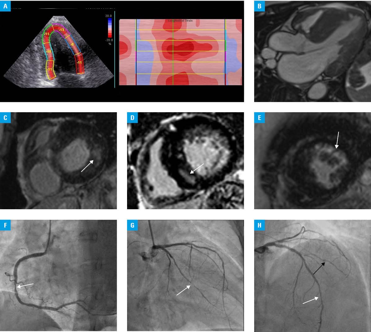
Cardiac magnetic resonance (CMR) confirmed concentric biventricular hypertrophy without systolic anterior motion of the mitral valve (Figure 1B). Stress perfusion imaging was performed during adenosine infusion (140 µg/kg/min), and perfusion assessment was qualitative. First‑pass perfusion imaging showed a regional perfusion defect predominantly in the basal inferoseptal segments (Figure 1C). Perfusion imaging in storage cardiomyopathies allows for differentiation between microvascular dysfunction and true ischemic injury.2
Native T1 mapping (Magnetic Occult Lesion Localization Instrument sequence, 1.5 T) demonstrated reduced global myocardial T1 values of 890 ms (reference range [RR], 950–1050 ms), with the lowest values observed in the basal inferolateral segments. The extracellular volume fraction was 24% (RR <28%), supporting predominant intracellular sphingolipid storage rather than diffuse interstitial fibrosis. Late gadolinium enhancement (LGE) demonstrated a mixed pattern of myocardial injury. Nonischemic mid‑wall LGE involved the inferolateral wall and basal interventricular septum (Figure 1D), typical of Fabry cardiomyopathy.3 Concurrently, subendocardial ischemic LGE was observed in mid‑to‑apical anterior and anterolateral segments (Figure 1E), suggesting coexisting epicardial coronary artery disease (CAD).4
Given the regional perfusion defect on stress CMR and a high pretest probability of epicardial CAD, additional nuclear perfusion imaging was deemed unlikely to alter clinical management. The patient was referred directly for invasive coronary angiography, which identified multivessel CAD, with significant stenoses in the right coronary artery (Figure 1F) and in the obtuse marginal and diagonal branches (Figure 1G and 1H), as well as a functionally significant lesion in the left anterior descending artery, confirmed by the diastolic hyperemia‑free ratio (0.5) and fractional flow reserve (0.78; RR ≥0.89; Figure 1H). After the case was discussed by the heart team, the patient was deemed unsuitable for coronary artery bypass grafting and subsequently referred for percutaneous coronary intervention.
This case emphasizes the importance of a comprehensive diagnostic approach in patients with Fabry disease, in whom myocardial ischemia is often attributed to microvascular dysfunction, while coexisting epicardial CAD may also be present, particularly in older individuals. Glycosphingolipid accumulation within endothelial and vascular smooth muscle cells leads to progressive coronary microvascular dysfunction, impaired coronary flow reserve, and reduced nitric oxide bioavailability, resulting in myocardial ischemia, even in the absence of obstructive epicardial CAD. Concentric LV hypertrophy further increases myocardial oxygen demand, aggravating ischemic imbalance. Recent data from Polish cohorts demonstrate substantial genotype–phenotype variability in patients screened for LV hypertrophy, further emphasizing the importance of precise genetic characterization.5 The diagnostic and therapeutic strategy applied in this case was consistent with the current European Society of Cardiology guidelines on cardiomyopathies (2023)6 and chronic coronary syndromes (2024),7 which support the use of multiparametric CMR for tissue characterization and functional ischemia assessment, as well as invasive coronary angiography in patients with documented regional ischemia and a high pretest probability of epicardial CAD. Multiparametric CMR, including stress perfusion and LGE imaging, enables accurate differentiation between ischemic and nonischemic myocardial injury, and guides appropriate clinical management.2-4
- Lu DY, Huang WM, Wang WT, et al. Reduced global longitudinal strain as a marker for early detection of Fabry cardiomyopathy. Eur Heart J Cardiovasc Imaging. 2022; 23: 487‑495. | Crossref
- Chau HHT, Fung WY, Ma JKF, et al. Cardiac magnetic resonance imaging in Fabry disease. J Clin Imaging Sci. 2025; 15: 23. | Crossref
- Pieroni M, Zocchi C, Ciabatti M. Cardiac involvement in Fabry disease: recent advances, unresolved issues, and unmet needs. Eur Heart J Suppl. 2025; 27: i51‑i55. | Crossref
- Li X, Ren X, Zhang Y, et al. Fabry disease: mechanism and therapeutics strategies. Front Pharmacol. 2022; 13: 1025740. | Crossref
- Szczepara S, Pacia K, Trojanowicz K, et al. Looking for Fabry, finding more: LVH screening yields unexpected Gaucher diagnosis. Med Sci (Basel). 2025; 13: 162. | Crossref
ARTICLE INFORMATION