

 CC BY 4.0
A 47‑year‑old man was admitted with a 1‑month history of recurrent cough, sputum production, and dyspnea. Intermittent yellow‑white sputum was reported without fever or hemoptysis. No significant medical history was documented. Laboratory investigations showed elevated leukocyte (10.66 × 10⁹/l; reference range [RR], 4–10 × 109/l) and neutrophil (7.67 × 10⁹/l; RR, 1.8–6.3 × 10⁹/l) counts. Chest computed tomography (Figure 1A–1D) demonstrated diffuse ground‑glass opacities in both lungs, with thickened interlobular and intralobular septa, presenting the characteristic “crazy‑paving” pattern. Bronchoscopic lung biopsy and whole‑lung lavage were performed under general anesthesia. Milky lavage fluid was obtained, and both tissue and fluid specimens showed positive periodic acid‑Schiff staining, confirming a diagnosis of pulmonary alveolar proteinosis (PAP) through demonstration of alveolar lipoproteinaceous deposits.
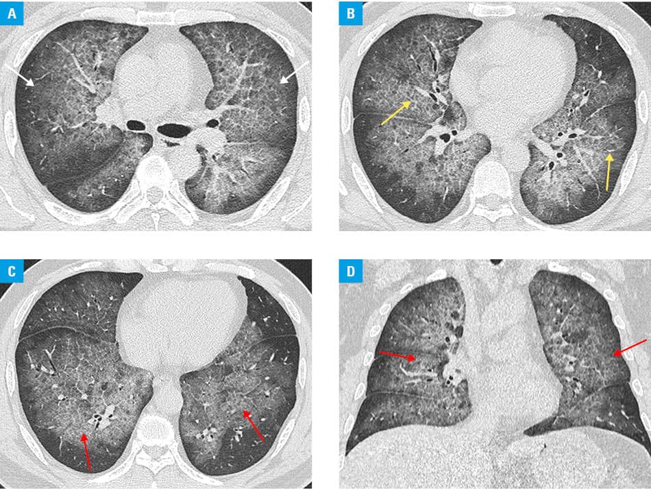
PAP is a rare lung disorder characterized by abnormal accumulation of surfactant material within alveolar spaces, impairing gas exchange. The disease is primarily localized in the lungs, with most patients exhibiting an insidious onset and symptoms, including dyspnea, cough, and occasional sputum production. Three variants are recognized: autoimmune, accounting for approximately 90% of the cases, and associated with anti–granulocyte‑macrophage colony‑stimulating factor antibodies1; secondary, resulting from hematologic malignancies, infections, or inhalational exposures; and hereditary, linked to mutations in genes involved in surfactant production. Saline‑based whole‑lung lavage remains the primary treatment.2 Although pulmonary function typically improves following lavage in autoimmune cases, median remission period of 15 months has been reported,3 potentially necessitating repeat procedures. In the current case, sequential bilateral lung lavage performed in 6 sessions resulted in symptomatic improvement and complete radiologic resolution.
- Bendstrup E, Lynn E, O’Callaghan M. Recent advances in the diagnosis and management of pulmonary alveolar proteinosis. Expert Rev Respir Med. 2025; 19: 1247‑1261. | Crossref
- Bonella F, Borie R. Pulmonary alveolar proteinosis. Clin Chest Med. 2025; 46: 633‑647. | Crossref
- McCarthy C, Bonella F, O’Callaghan M, et al. European Respiratory Society guidelines for the diagnosis and management of pulmonary alveolar proteinosis. Eur Respir J. 2024; 64: 2400725. | Crossref
ARTICLE INFORMATION